|
|
Welcome to The Visible Embryo, a comprehensive educational resource on human development from conception to birth.
The Visible Embryo provides visual references for changes in fetal development throughout pregnancy and can be navigated via fetal development or maternal changes.
The National Institutes of Child Health and Human Development awarded Phase I and Phase II Small Business Innovative Research Grants to develop The Visible Embryo. Initally designed to evaluate the internet as a teaching tool for first year medical students, The Visible Embryo is linked to over 600 educational institutions and is viewed by more than one million visitors each month.
Today, The Visible Embryo is linked to over 600 educational institutions and is viewed by more than 1 million visitors each month. The field of early embryology has grown to include the identification of the stem cell as not only critical to organogenesis in the embryo, but equally critical to organ function and repair in the adult human. The identification and understanding of genetic malfunction, inflammatory responses, and the progression in chronic disease, begins with a grounding in primary cellular and systemic functions manifested in the study of the early embryo.

The World Health Organization (WHO) has created a new Web site to help researchers, doctors and
patients obtain reliable information on high-quality clinical trials. Now you can go to one website and search all registers to identify clinical trial research underway around the world!

|
|
| Disclaimer: The Visible Embryo web site is provided for your general information only. The information contained on this site should not be treated as a substitute for medical, legal or other professional advice. Neither is The Visible Embryo responsible or liable for the contents of any websites of third parties which are listed on this site. |
|
|
 |
|
Content protected under a Creative Commons License. Commons License.
No dirivative works may be made or used for commercial purposes. |
|
|
| |
|
|

CLICK ON weeks 0 - 40 and follow along every 2 weeks of fetal development
|
|
|
|
|
Home | Pregnancy Timeline | News Alerts |News Archive Nov 19, 2014
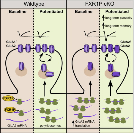
The protein, FXR1P (Fragile X Related Protein 1), suppresses production of molecules required
for building memories. When FXR1P is selectively removed from certain parts of the brain, new
molecules are produced that strengthened connections between brain cells and correlates
with improved memory and recall in mice.
Image credit: Elsevier Inc. |
|
|
|
|
|
The science behind Total Memory Recall
Is it possible to change the amount of information the brain can store? Maybe, according to a new international study led by the Research Institute of the McGill University Health Centre (RI-MUHC).
The research has identified a molecule that puts a brake on brain processing. When the brake is removed, brain function and memory recall is improved.
Published in the latest issue of Cell Reports the study has implications for neurodevelopmental and neurodegenerative diseases, such as autism spectral disorders and Alzheimer’s.
“Previous research has shown that production of new molecules is necessary for storing memories in the brain; if you block the production of these molecules, new memory formation does not take place. Our findings show that the brain has a key protein that limits the production of molecules necessary for memory formation. When this brake-protein is suppressed, the brain is able to store more information.”
Dr. Keith Murai, RI-MUHC neuroscientist, study senior author and Associate Professor in the Department of Neurology and Neurosurgery at McGill University.
The researchers used a mouse model to study how changes in brain cell connections produce new memories.
Dr. Murai and his colleagues found that the protein, FXR1P (Fragile X Related Protein 1), is responsible for suppressing production of molecules required for building new memories.
When FXR1P is selectively removed from certain parts of the brain, new molecules are produced that strengthen connections between brain cells. This result correlates with improved memory and recall in mice.
Dr. Murai: “The role of FXR1P was a surprising discovery. Previous to our work, no-one had identified a role for this regulator in the brain. Our findings have provided fundamental knowledge about how the brain processes information. We’ve identified a new pathway that directly regulates how information is handled and could have relevance for understanding and treating brain diseases.”
“Future research in this area could be very interesting. If we can identify compounds that control the braking potential of FXR1P, we may be able to alter amounts of brain activity or plasticity. For example, in autism, we may want to decrease certain brain activity — but in Alzheimer’s, we may want to enhance activity. By manipulating FXR1P, we may eventually be able to adjust memory formation and retrieval, thus improving the quality of life of people suffering from brain diseases.”
Highlights
•Removal of FXR1P increases protein synthesis-dependent L-LTP and memory storage
•FXR1P limits GluA2 synthesis and its activity-dependent synaptic delivery
•FXR1P represses GluA2 mRNA translation via a GU-rich element in its 5′ UTR
•Fragile X proteins have divergent roles in synaptic plasticity and memory storage
Summary
Translational control of mRNAs allows for rapid and selective changes in synaptic protein expression that are required for long-lasting plasticity and memory formation in the brain. Fragile X Related Protein 1 (FXR1P) is an RNA-binding protein that controls mRNA translation in nonneuronal cells and colocalizes with translational machinery in neurons. However, its neuronal mRNA targets and role in the brain are unknown. Here, we demonstrate that removal of FXR1P from the forebrain of postnatal mice selectively enhances long-term storage of spatial memories, hippocampal late-phase long-term potentiation (L-LTP), and de novo GluA2 synthesis. Furthermore, FXR1P binds specifically to the 5′ UTR of GluA2 mRNA to repress translation and limit the amount of GluA2 that is incorporated at potentiated synapses. This study uncovers a mechanism for regulating long-lasting synaptic plasticity and spatial memory formation and reveals an unexpected divergent role of FXR1P among Fragile X proteins in brain plasticity.
This is an open access article under the CC BY-NC-ND license (http://creativecommons.org/licenses/by-nc-nd/3.0/).
This research was made possible with funding from the Canadian Institutes of Health Research (CIHR), the Natural Sciences and Engineering Research Council of Canada, and National Institutes of Health (U.S.A.).
Return to top of page |