|
|
Mitochondria divide differently than once thought
For the first time a study reveals how mitochondria, the power generators found in nearly all living cells, regularly divide and multiply.
In 2011, the University of Colorado Boulder (CU Boulder) Gia Voeltz PhD, Associate Professor, along with her team, found that the organelle structure known as the endoplasmic reticulum (ER) branches throughout the cell cytoplasm like a spider web. ER wraps around other cell organelles — including mitochondria, and once an ER tentacle touches a mitochondrion — that mitochondrion (1) begins to constrict. Then another cell protein — Dynamin-related protein (Drp1) — is recruited to (2) help constriction at the original point of contact.
Voeltz's team then found that once the squeeze of mitochondria by Drp1 begins, a second protein called Dynamin-2, or Dyn2, is recruited to (3) finish the job in a process called fission, splitting the mitochondrion into two.
Shaped like tiny springs, Dynamin proteins are enlisted by the endoplasmic reticulum to encircle mitochondria — squeezing and twisting its elongated shape into two separate organelles.
Both proteins are needed as Drp1 is only strong enough to squeeze the mitochondria fractionally in a process that Dyn2 must finish.
"Our findings change what everyone has believed about mitochondrial division," said postdoctoral fellow Jason Lee, first author on the study. "Now we know that it takes at least three different constriction steps in order to ultimately divide mitochondria."
The paper was published in Nature Oct. 31, 2016.
Floating in almost all living cells, mitochondria vary in number from dozens to several thousand. For example, muscle cells have large numbers of mitochondria because of their high energy needs.
New mitochondria are created when cells signal their need for more energy.
Mitochondria play a role in longevity, too. They are crucial for blood sugar maintenance and fat loss. Damaged mitochondria can cause problems in the brain, liver, heart, skeletal muscles and respiratory systems. Knowing how their division process works brings scientists a step closer to understanding what process might be changing under pathological conditions like cancer.
Jason Lee: "The ability of our cells to efficiently convert nutrients into energy is rooted in a cell's ability to manage mitochondria shape, number and position through a balance of fusion and division. This balance goes awry in cancer and neurodegeneration."
Abstract
Mitochondria cannot be generated de novo; they must grow, replicate their genome, and divide in order to be inherited to each daughter cell during mitosis. Mitochondrial division is a structural challenge that requires a massive remodelling of membrane morphology1,2,3. Although division factors differ across organisms, the need for multiple constriction steps and a dynamin-related protein (Drp1, Dnm1 in yeast) has been conserved4,5,6. In mammalian cells, mitochondrial division has been shown to proceed with at least two sequential constriction steps: (1) endoplasmic reticulum (ER) and actin collaborate to generate constrictions suitable for Drp1 assembly; (2) Drp1 further constricts membranes until fission occurs2,7,8,9. However, in vitro experiments argue that Drp1 does not have the dynamic range to complete membrane fission per se7. In contrast to Drp1, the neuronal-specific classical dynamin-1 (Dyn1) has been shown to assemble on narrower lipid profiles and facilitates spontaneous membrane fission upon GTP hydrolysis10,11. Here we discovered that the ubiquitously expressed classical dynamin-2 (Dyn2) is a fundamental component of the mitochondrial division machinery. A combination of live-cell and electron microscopy reveals that Dyn2 works in concert with Drp1 to orchestrate sequential constriction events leading up to division. Our work underscores the biophysical limitations of Drp1 and positions Dyn2, which has intrinsic membrane fission properties, at the final step of mitochondrial division.
In addition to Voeltz and Lee, other CU Boulder paper contributors included postdoctoral fellow Laura Westrate, graduate student Haoxi Wu and researcher Cynthia Page. All study authors are in the Department of Molecular, Cellular and Developmental Biology.
The new study was funded by grants from the National Institutes of Health.
Return to top of page
|
|
|
Nov 2, 2016 Fetal Timeline Maternal Timeline News News Archive
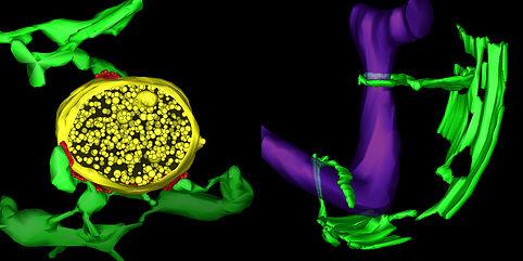
The electron tomograph above reveals the three-dimensional structure of membrane contact sites (RED)
between Endoplasmic Reticulum tubules (GREEN) and mitochondria (PURPLE) that are found in a yeast cell on the right; and an endosome (YELLOW) in an animal cell on the left.
Image Credit: Matthew West.
________________________
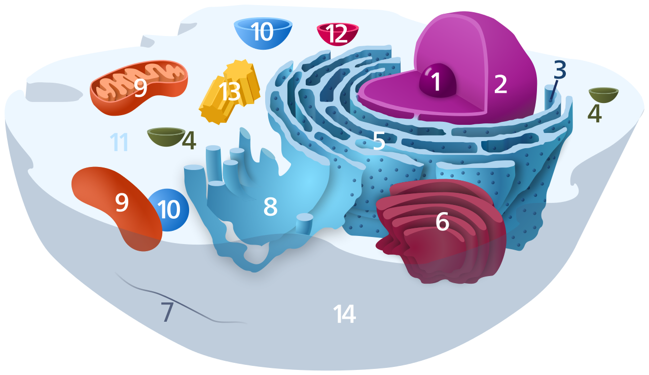
BELOW: A diagram of an animal cell and its parts in 2 dimensions — 1. Nucleolus, 2. Nucleus, 3. Ribosome, 4. Vesicle, 5. Rough endoplasmic reticulum, 6. Golgi apparatus (or "Golgi body"), 7. Cytoskeleton, 8. Smooth endoplasmic reticulum, 9. Mitochondrion, 10. Vacuole, 11. Cytosol, 12. Lysosome, 13. Centriole, 14. Cell Wall Image Credit: Wikipedia
|


