|

CLICK ON weeks 0 - 40 and follow along every 2 weeks of fetal development
|
||||||||||||||||||||||||||||
Gene therapy restores hearing and balance In France, one child in 700 is born with severe or profound hearing loss, and one in every 1,000 will lose their sense of hearing before adulthood. Hearing loss, sometimes associated with other disorders such as a balance defect, is the most common sensory deficit affecting more than 280 million people worldwide, according to the World Health Organization or WHO. Over the past 20 years, scientists have made remarkable progress in deciphering the genetic origins of congenital hereditary hearing loss, which is usually caused by inner ear dysfunction. The inner ear is made up of: the hearing organ or cochlea five balance organs 1) the saccule 2) utricle with 3) 4) 5) being three semicircular canals containing sensory cells, or hair cells which detect mechanical vibrations and convert them into electrical signals. 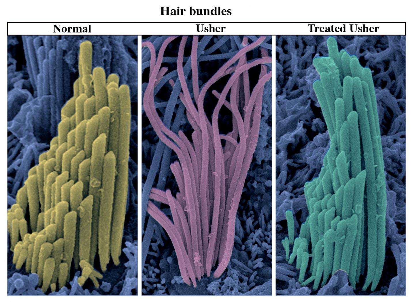 Mutations in more than 100 genes have been associated with inner ear defects causing deafness. The various hereditary forms of hearing loss include Usher syndrome type 1 (USH1), a particularly severe clinical form of deaf-blindness, specifically the USH1G genetic form. USH1G patients are profoundly deaf and have no balance function at birth. They subsequently suffer from prepubertal-onset sight loss leading to blindness. USH1G syndrome is due to mutations in the gene encoding the scaffold protein sans, essential for cohesion of the hair bundle within inner ear hair cells. Patients with hearing loss and balance dysfunction are currently fitted with auditory "hearing aids" and may be given balance rehabilitation therapy. But, the outcomes are variable. One possible alternative for treating hereditary inner ear defects is gene therapy. This approach involves transferring a non-mutated copy of the already defective gene to restore the missing protein. So far, gene therapy attempts have only partially improved hearing in mouse models of specific human deafness types not including severe anomalies in hair cell structure. But now, scientists from the Institut Pasteur, Inserm, the CNRS, Collège de France, University Pierre et Marie Curie, and University Clermont Auvergne*, have succeeded in restoring hearing and balance in a mouse model of USH1G syndrome using gene therapy. With a single local injection of the USH1G gene just after birth, the scientists observed restoration of the structure and mechanosensory function of the inner ear hair bundles - profoundly damaged before birth - which resulted in (1) long-term partial recovery of hearing, and (2) complete recovery of vestibular balance in these mice. These results unexpectedly establish that inner ear defects due to major morphogenetic abnormalities of the hair bundle can be reversed even after birth by gene therapy. Scientists injected the USH1G gene into the inner ear using an innocuous AAV8 virus, which enabled them to target hair cells. Expression of the therapeutic gene was detected 48 hours after injection. The team demonstrated that a single injection to restore the production and localization of the missing protein in hair cells successfully improved hearing and balance functions in young mice. These findings suggest the therapeutic protein was able to interact normally with its binding partners (proteins cadherin 23, protocadherin 15, myosin VIIA and harmonin) in the mechano-electrical transduction to the hair bundles. "We have just shown that it is possible to partially correct a specific form of hereditary hearing loss, accompanied by balance problems, using local gene therapy performed after embryogenesis of the ear which is primarily affected by the mutation responsible for the disorder. The study is published in the Proceedings of the National Academy of Sciences (PNAS) and represents a significant step towards clinical trials in gene therapy for the curative treatment of hereditary deafness and balance loss in humans. Significance Hearing and balance impairments are major concerns and a serious burden for public health, but still lack an effective curative therapy. We assessed inner ear functions in a mouse model of Usher syndrome type 1, a developmental disorder characterized by profound congenital deafness and balance deficit, after local gene therapy. Viral transfer of the wild-type cDNA to the inner ear of the mutant mice shortly after birth resulted in a partial restoration of hearing and a long-lasting, almost complete, removal of the balance defect. The present results provide a basis for future clinical trials in humans. Abstract Our understanding of the mechanisms underlying inherited forms of inner ear deficits has considerably improved during the past 20 y, but we are still far from curative treatments. We investigated gene replacement as a strategy for restoring inner ear functions in a mouse model of Usher syndrome type 1G, characterized by congenital profound deafness and balance disorders. These mice lack the scaffold protein sans, which is involved both in the morphogenesis of the stereociliary bundle, the sensory antenna of inner ear hair cells, and in the mechanoelectrical transduction process. We show that a single delivery of the sans cDNA by the adenoassociated virus 8 to the inner ear of newborn mutant mice reestablishes the expression and targeting of the protein to the tips of stereocilia. The therapeutic gene restores the architecture and mechanosensitivity of stereociliary bundles, improves hearing thresholds, and durably rescues these mice from the balance defects. Our results open up new perspectives for efficient gene therapy of cochlear and vestibular disorders by showing that even severe dysmorphogenesis of stereociliary bundles can be corrected. Authors: Alice Emptoza, Vincent Michel, Andrea Lelli, Omar Akild, Jacques Boutet de Monvel, Ghizlene Lahlou, Anaïs Meyer, Typhaine Dupont, Sylvie Nouaille, Elody Eye, Filipa Franca de Barros, Mathieu Beraneck, Didier Dulong, Jean-Pierre Hardelin, Lawrence Lustigh, Paul Avani, Christine Petit, and Saaid Safieddine *From the Genetics & Physiology of Hearing Laboratory (Institut Pasteur/Inserm/UPMC), the Genes, Synapses and Cognition Laboratory (CNRS/Institut Pasteur, the Center for Neurophysics, Physiology and Pathology (CNRS/Paris-Descartes University), and the Sensory Biophysics Laboratory (University Clermont Auvergne). Return to top of page |
Oct 31, 2017 Fetal Timeline Maternal Timeline News News Archive 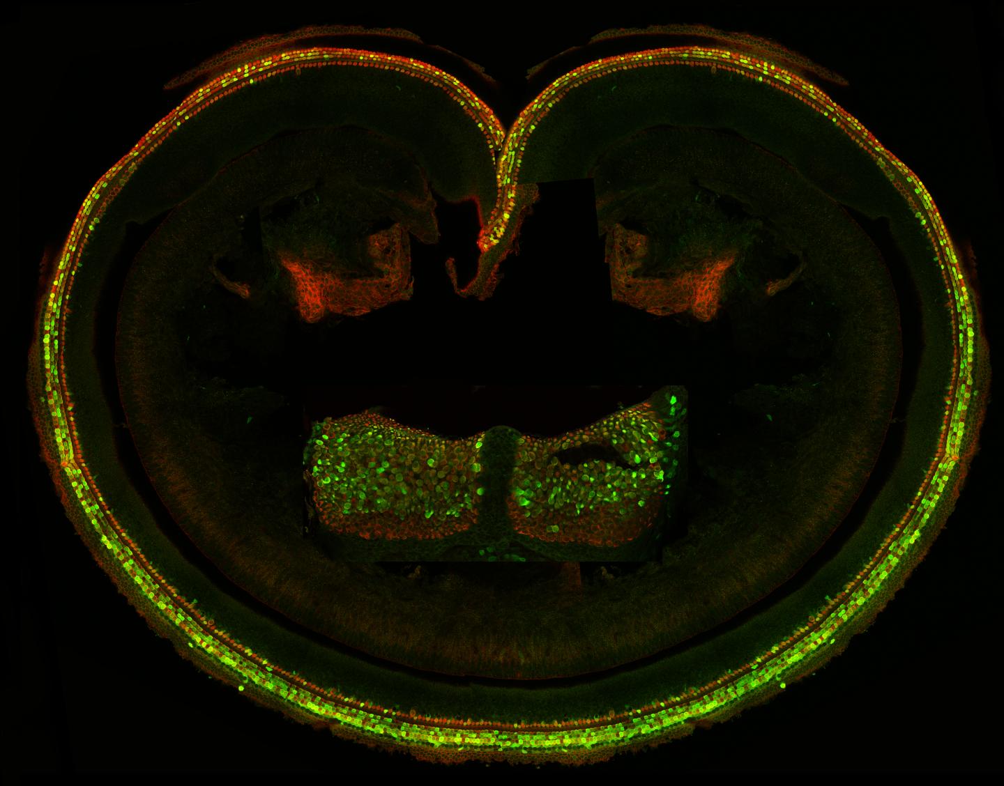 Confocal microscope image of cochlear sensory epithelium (organ of Corti) of the inner ear. Green and orange cells produce proteins from the transferred therapeutic gene, introduced through the AAV8 virus.
|
||||||||||||||||||||||||||||

