Welcome to The Visible Embryo
  o
 
The Visible Embryo Birth Spiral Navigation
   
Google  
Fetal Timeline--- -Maternal Timeline-----News-----Prescription Drugs in Pregnancy---- Pregnancy Calculator----Female Reproductive System

   
WHO International Clinical Trials Registry Platform

The World Health Organization (WHO) has a Web site to help researchers, doctors and patients obtain information on clinical trials.

Now you can search all such registers to identify clinical trial research around the world!


Home

History

Bibliography

Pregnancy Timeline

Prescription Drug Effects on Pregnancy

Pregnancy Calculator

Female Reproductive System

News

Disclaimer: The Visible Embryo web site is provided for your general information only. The information contained on this site should not be treated as a substitute for medical, legal or other professional advice. Neither is The Visible Embryo responsible or liable for the contents of any websites of third parties which are listed on this site.


Content protected under a Creative Commons License.
No dirivative works may be made or used for commercial purposes.


Pregnancy Timeline by SemestersDevelopmental TimelineFertilizationFirst TrimesterSecond TrimesterThird TrimesterFirst Thin Layer of Skin AppearsEnd of Embryonic PeriodEnd of Embryonic PeriodFemale Reproductive SystemBeginning Cerebral HemispheresA Four Chambered HeartFirst Detectable Brain WavesThe Appearance of SomitesBasic Brain Structure in PlaceHeartbeat can be detectedHeartbeat can be detectedFinger and toe prints appearFinger and toe prints appearFetal sexual organs visibleBrown fat surrounds lymphatic systemBone marrow starts making blood cellsBone marrow starts making blood cellsInner Ear Bones HardenSensory brain waves begin to activateSensory brain waves begin to activateFetal liver is producing blood cellsBrain convolutions beginBrain convolutions beginImmune system beginningWhite fat begins to be madeHead may position into pelvisWhite fat begins to be madePeriod of rapid brain growthFull TermHead may position into pelvisImmune system beginningLungs begin to produce surfactant
CLICK ON weeks 0 - 40 and follow along every 2 weeks of fetal development


Developmental biology - Cell Division

Why do we need a pair of genome?

Unraveling how cell division destabilizes a cell with more — or less — than one pair of chromosomes...


Scientists have unraveled how the replication of a cell destabilizes that cell when it has more, or less, than a pair of chromosome sets - each called a genome. This is a major step towards understanding chromosome instability in cancer cells.

In mammals, including humans, cells that make up the body are diploids as each cell has a pair of chromosomes. A non-diploid state destabilizes a cell, triggering abnormalities, cancers and other serious diseases.

In asexual reproduction and cancer, haploid (one set of each chromosome) and tetraploid (four sets of each chromosome) cells are created which causes chromosomes to become unstable. However, why such instability occurs when a cell is in non-diploid isn't understood.

In the present study, published in the Journal of Cell Biology, researchers used human cell lines in different states - haploid, diploid and tetraploid - to investigate the effect different chromosome configurations have on cell replication.
In normal cells there are two centrosomes that regulate cell replication. Researchers saw that gradual loss of centrosomes in haploid cells, and frequent duplication of centrosomes in tetraploid cells, trigger abnormalities in cell replication.

In addition, they found fewer cell fibers — microtubules — in haploid cells, but more microtubules in tetraploid cells. This is significant as the number of microtubules changes the efficiency of centrosome duplication. This results in either loss of the centrosome or it's over-duplication.

Of interest is that the efficiency of DNA replication, remained constant regardless of over-duplication of the centrosome.

"Incompatibility between the centrosome duplication and the DNA replication cycle could be the underlying cause of the instability in non-diploid cells - such as cancer cells - in mammals. Our findings could help us understand chromosome instability in cancer cells, which are often in a non-diploid state, and lead to new cancer treatment strategies." explains Ryota Uehara, PhD.


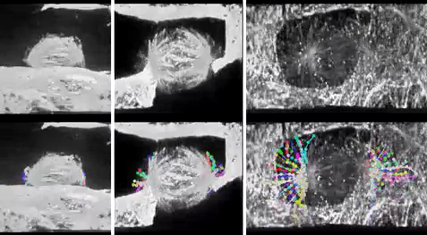
Scene from Video 7: 3D reconstruction of HAP1 cells.


Abstract
In animals, somatic cells are usually diploid and are unstable when haploid for unknown reasons. In this study, by comparing isogenic human cell lines with different ploidies, we found frequent centrosome loss specifically in the haploid state, which profoundly contributed to haploid instability through subsequent mitotic defects. We also found that the efficiency of centriole licensing and duplication changes proportionally to ploidy level, whereas that of DNA replication stays constant. This caused gradual loss or frequent overduplication of centrioles in haploid and tetraploid cells, respectively. Centriole licensing efficiency seemed to be modulated by astral microtubules, whose development scaled with ploidy level, and artificial enhancement of aster formation in haploid cells restored centriole licensing efficiency to diploid levels. The ploidy–centrosome link was observed in different mammalian cell types. We propose that incompatibility between the centrosome duplication and DNA replication cycles arising from different scaling properties of these bioprocesses upon ploidy changes underlies the instability of non-diploid somatic cells in mammals.

Authors: Kan Yaguchi, Takahiro Yamamoto, Ryo Matsui, View ORCID ProfileYuki Tsukada, Atsuko Shibanuma, Keiko Kamimura, Toshiaki Koda, Ryota Uehara.


Acknowledgements
This research was supported by the UC San Francisco California Preterm Birth Initiative, which is funded by Marc and Lynne Benioff. Additional support was provided by grants from the National Institute of Environmental Health Sciences (K99ES027023, P01ES022841, R01ES027051) and the U.S. Environmental Protection Agency (RD-83543301).

Return to top of page

Jun 14, 2018   Fetal Timeline   Maternal Timeline   News   News Archive


A diploid cell (LEFT) and A haploid cell (RIGHT) showing normal and abnormal orientation
of chromosomes (PURPLE) with microtubules (GREEN) during cell division.
Image credit: Yaguchi K., et al., Journal of Cell Biology.


Phospholid by Wikipedia