Welcome to The Visible Embryo
  o
 
The Visible Embryo Birth Spiral Navigation
   
Google  
Fetal Timeline--- -Maternal Timeline-----News-----Prescription Drugs in Pregnancy---- Pregnancy Calculator----Female Reproductive System

   
WHO International Clinical Trials Registry Platform

The World Health Organization (WHO) has a Web site to help researchers, doctors and patients obtain information on clinical trials.

Now you can search all such registers to identify clinical trial research around the world!


Home

History

Bibliography

Pregnancy Timeline

Prescription Drug Effects on Pregnancy

Pregnancy Calculator

Female Reproductive System

News

Disclaimer: The Visible Embryo web site is provided for your general information only. The information contained on this site should not be treated as a substitute for medical, legal or other professional advice. Neither is The Visible Embryo responsible or liable for the contents of any websites of third parties which are listed on this site.


Content protected under a Creative Commons License.
No dirivative works may be made or used for commercial purposes.


Pregnancy Timeline by SemestersDevelopmental TimelineFertilizationFirst TrimesterSecond TrimesterThird TrimesterFirst Thin Layer of Skin AppearsEnd of Embryonic PeriodEnd of Embryonic PeriodFemale Reproductive SystemBeginning Cerebral HemispheresA Four Chambered HeartFirst Detectable Brain WavesThe Appearance of SomitesBasic Brain Structure in PlaceHeartbeat can be detectedHeartbeat can be detectedFinger and toe prints appearFinger and toe prints appearFetal sexual organs visibleBrown fat surrounds lymphatic systemBone marrow starts making blood cellsBone marrow starts making blood cellsInner Ear Bones HardenSensory brain waves begin to activateSensory brain waves begin to activateFetal liver is producing blood cellsBrain convolutions beginBrain convolutions beginImmune system beginningWhite fat begins to be madeHead may position into pelvisWhite fat begins to be madePeriod of rapid brain growthFull TermHead may position into pelvisImmune system beginningLungs begin to produce surfactant
CLICK ON weeks 0 - 40 and follow along every 2 weeks of fetal development


Developmental Biology - Precision Medicine

Regrowing Damaged Kidneys

Scientists discover newborns have ability to repair their kidneys...


Scientists seeking to regrow kidneys make a promising discovery — kidneys can repair themselves immediately following birth. Newborns have a remarkable ability to self repair after a kidney obstruction is removed. This finding offers insights into how repair happens and might eventually help doctors regenerate kidneys in adults.

Research conducted at the University of Virginia School of Medicine (UVA), was looking at the effects of surgically removing kidney blockages in newborn mice, when they made the observation. Their findings stress the importance of speedy surgical intervention after human births as well, according to Maria Luisa S. Sequeira-Lopez MD, at UVA's Child Health Research Center. The research is published in the journal Clinical Science.

Dr. Sequeira-Lopez described another strange phenomenon seen in blocked kidneys: "When you obstruct the kidney, and keep it obstructed, its vasculature shrinks. However, the other unobstructed kidney grows many more branches — as if trying to compensate," she explains. "Releasing the obstruction, the vasculature [vessels] of the previously obstructed kidney regenerates and grows dramatically — initiating regeneration of the whole kidney."

Preventing Kidney Damage

There are many reasons why a baby might be born with obstructed kidneys, known as "obstructive nephropathy." For example, its urinary tract might not be fully developed, or there could be a mass compressing it, which can cause serious problems including kidney failure.

Dr. Sequeira-Lopez: "When [the urinary tract] is blocked, it puts backward pressure on the kidney causing swelling, called hydronephrosis. When this happens, all the nephron tubules become damaged and eventually are lost. If hydronephrosis persists for a long time, it leads to end-stage renal disease."

Researchers were surprised at how quickly and effectively damaged kidneys of newborn mice repair. They identified "precursor cells" as playing a critical role in that process and hope to harness the human version of these cells to perform similar kidney repairs in both children and adults.
"During development, there are more precursor cells. Studying them may give us clues on how to push these cells to come awake in the adult and regenerate to maintain kidney function."

"Moving forward, we want to focus on how kidney vasculature regenerates... because it's very, very important to know all mechanisms of cellular and molecular change...not only relevant for congenital nephropathy alone, it could apply to all kidney disease."


Maria Luisa S. Sequeira-Lopez MD, Child Health Research Center, University of Virginia School of Medicine, Charlottesville, Virginia, USA.

Abstract
Congenital obstructive nephropathy is a major cause of chronic kidney disease (CKD) in children. The contribution of changes in the identity of renal cells to the pathology of obstructive nephropathy is poorly understood. Using a partial unilateral ureteral obstruction (pUUO) model in genetically modified neonatal mice, we traced the fate of cells derived from the renal stroma, cap mesenchyme, ureteric bud (UB) epithelium, and podocytes using Foxd1Cre, Six2Cre, HoxB7Cre, and Podocyte.Cre mice respectively, crossed with double fluorescent reporter (membrane-targetted tandem dimer Tomato (mT)/membrane-targetted GFP (mG)) mice. Persistent obstruction leads to a significant loss of tubular epithelium, rarefaction of the renal vasculature, and decreased renal blood flow (RBF). In addition, Forkhead Box D1 (Foxd1)-derived pericytes significantly expanded in the interstitial space, acquiring a myofibroblast phenotype. Degeneration of Sine Oculis Homeobox Homolog 2 (Six2) and HoxB7-derived cells resulted in significant loss of glomeruli, nephron tubules, and collecting ducts. Surgical release of obstruction resulted in striking regeneration of tubules, arterioles, interstitium accompanied by an increase in blood flow to the level of sham animals. Contralateral kidneys with remarkable compensatory response to kidney injury showed an increase in density of arteriolar branches. Deciphering the mechanisms involved in kidney repair and regeneration post relief of obstruction has potential therapeutic implications for infants and children and the growing number of adults suffering from CKD.

Authors
Nagalakshmi, Minghong Li, Soham Shah, Joseph C. Gigliotti, Alexander L. Klibanov, Frederick H. Epstein, Robert L. Chevalier, R. Ariel Gomez and Sequeira-Lopez.

Acknowledgements
The work was supported by the National Institutes of Health, grants DK091330, DK096373 and DK116196.

About the Journal: Clinical Science
Translating molecular bioscience and experimental research into medical insights, Clinical Science offers multi-disciplinary coverage and clinical perspectives to advance human health. In addition to original papers, the journal features state-of-the-art review articles, hypotheses, invited commentaries and scientific correspondence on recently published papers appearing in the journal. Translating molecular bioscience and experimental research into medical insights, Clinical Science offers multi-disciplinary coverage and clinical perspectives to advance human health.

Return to top of page

Feb 11, 2019   Fetal Timeline   Maternal Timeline   News  
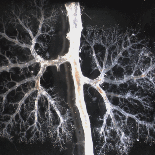


Obstructive nephropathy is the leading identifiable cause of renal failure in children. However,
current management is limited to surgical removal of the obstruction, which often fails to prevent
progressive tissue damage. UVA research investigates the neonatal mouse, which parallels
urinary tract obstruction in the human fetus. Image: Fetal kidney branching, UVA.


Phospholid by Wikipedia