|
|
Developmental Biology - Quantum Biology
A Circuit Board In Each Cell?
Are cells wired like a computer chip, directing signals and instructing function?...
Research suggests cells in our body are wired like computer chips and receive and direct signals instructing them how to function. However, unlike a fixed circuit board our cells can rapidly rewire their communication networks to change behaviour. The discovery of this cell wide web turns on its head, our understanding of how instructions spread inside a cell.
It is thought the various structures inside a cell float around in an open sea called the cytoplasm. It is thought cellular signals tell the cell what to do and are transmitted in waves whose varied frequency is crucial to the message. Instead, researchers at the University of Edinburgh believe information is carried across a web of guide wires that transmit signals across nanoscale distances, just as found on a computer microprocessor.
They made their observations studying the movement of charged calcium molecules within cells using high-powered microscopes and computing techniques similar to those capturing the first ever image of a black hole in space.
Quantum Biology is an emerging field using quantum mechanics and theoretical chemistry to solve biological problems.
Thus localized signals are responsible for orchestrating cell activity which can relax or contract muscle cells. Signals to the nucleus, instruct minute changes releasing genes to be expressed (function). Change in gene expression is understood to alter cell behavior. When a cell moves from a steady state into its growth phase, signals switch on growth genes.
Researchers believe understanding the code controlling this wiring system could help us understand diseases such as pulmonary hypertension and cancer, and could one day open up new treatments. The study, published in Nature Communications, was funded by the British Heart Foundation.
"We found that cell function is coordinated by a network of nanotubes, similar to the carbon nanotubes you find in a computer microprocessor.
A most striking thing is this circuit is highly flexible. This cell-wide web can rapidly reconfigure delivery of different outputs determined by information received and relayed from the nucleus. Something no man-made microprocessors or circuit boards are yet capable of."
A. Mark Evans PhD, Professor, Centres for Discovery Brain Sciences and Cardiovascular Sciences, College of Medicine and Veterinary Medicine, University of Edinburgh, Scotland, UK.
Abstract
Ca2+ coordinates diverse cellular processes, yet how function-specific signals arise is enigmatic. We describe a cell-wide network of distinct cytoplasmic nanocourses with the nucleus at its centre, demarcated by sarcoplasmic reticulum (SR) junctions (<=400 nm across) that restrict Ca2+ diffusion and by nanocourse-specific Ca2+-pumps that facilitate signal segregation. Ryanodine receptor subtype 1 (RyR1) supports relaxation of arterial myocytes by unloading Ca2+ into peripheral nanocourses delimited by plasmalemma-SR junctions, fed by sarco/endoplasmic reticulum Ca2+ ATPase 2b (SERCA2b). Conversely, stimulus-specified increases in Ca2+ flux through RyR2/3 clusters selects for rapid propagation of Ca2+ signals throughout deeper extraperinuclear nanocourses and thus myocyte contraction. Nuclear envelope invaginations incorporating SERCA1 in their outer nuclear membranes demarcate further diverse networks of cytoplasmic nanocourses that receive Ca2+ signals through discrete RyR1 clusters, impacting gene expression through epigenetic marks segregated by their associated invaginations. Critically, this circuit is not hardwired and remodels for different outputs during cell proliferation.
Authors
Jingxian Duan, Jorge Navarro-Dorado, Jill H. Clark, Nicholas P. Kinnear, Peter Meinke, Eric C. Schirmer and A. Mark Evans.
Acknowledgements
Funding for this work was from a U.K. British Heart Foundation Programmed Grant (RG/12/14/29885; renewal rejected, December 2018) which supported the work of J.N.-D., J.D. and A.M.E. Microscopy was done in the IMPACT Imaging Facility at the University of Edinburgh. The work of J.D. also received support from the China Scholarship Council (201508060127). J.H.C. (PG/03/065) and N.P.K. (PG/05/128/19884) were supported by British Heart Foundation project grants to A.M.E. that expired in 2006 and 2007. E.C.S. and P.M. were supported by Wellcome grant 095209 and the Wellcome Centre for Cell Biology core grant 092076. J.D. and A.M.E. thank Heather McClafferty for support with respect to qPCR. The authors thank Sidney Fleischer, Frank Wuytack and Glenn Morris for the kind gift of antibodies. Finally, A.M.E. thanks Gordon Murray, Barclay Thomson and other members of the shortest day club for enlightening discussions on quantum tunnelling, and what might be possible when all the ducks are in a row.
Return to top of page
| |
|
May 28 2019 Fetal Timeline Maternal Timeline News
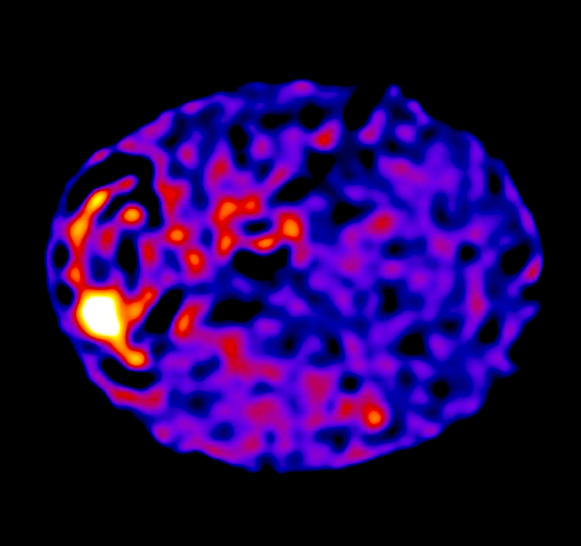 The first ever images of the cell-wide web have been captured by scientists at the University of Edinburgh. Their findings reveal cells in the body are wired like computer chips, directing signals that instruct function. Unlike a fixed circuit board, however, cells can rapidly rewire their communication networks and change their behavior. CREDIT The University of Edinburgh.
|


