|
|
Developmental Biology - Limb Formation
How Oxygen Shapes Arms and Legs
Origin of a mechanism called 'interdigital cell death'...
Amphibians, such as frogs and newts, form their limbs and toes by differential growth rates between digital and interdigital regions of their limbs.
Similarly, amniotes such as birds and mammals also employ cell death to shape their limbs. Removing parts of limb tissue through cell death allows for evolution in limb shapes — the lobed fingers of coots, even removal of some fingers in horses and camels. But, how all this works was unknown.
Researchers now have identified a surprising element that may be crucial to the appearance of interdigital cell death in four-footed animals ( tetrapods) during evolution: the amount of oxygen surrounding the embryo.
How can oxygen shape limbs?
The study, published in Developmental Cell, is a collaboration between three teams led by professors Mikiko Tanaka of Tokyo Tech, Haruki Ochi from Yamagata University and James Hanken of Harvard University.
Their goal was to understand the role of environmental oxygen in the evolution of four limbs.
Graduate student Ingrid Cordeiro and her colleagues used African clawed frogs (Xenopus laevis) as their model animal. This amphibian has interdigital membranes between the digits on its legs. When tadpoles are kept under high oxygen levels, some cells in these interdigital regions die, but, not in other body parts. Increasing the amount of blood vessels - the source by which oxygen reaches these interdigital regions - produced the same results showing how environmental change affects tadpole development in a very specific way.
Production of reactive oxygen species (ROS) is key to this process. ROS, often seen as detrimental to health, increases with aging and infertility.
However, researchers found ROS is not always bad, but a natural part of metabolism and can serve as a signal to cells during embryo formation. They found production of ROS in interdigital cell death is required in birds. Combined with previous reports in mammals, it is now possible to hypothesize the ROS mechanism is shared by all amniotes, including humans. However, some amphibians like the African clawed-frog and Japanese fire-bellied newt, have low ROS production in their interdigital regions.
Amphibian ecology and the evolution of limbs
Many amphibians are aquatic for part of their lives, breathing dissolved oxygen from water. In contrast amniotes, such as chickens, develop in an egg lined with membranes full of blood vessels. Gas exchange through more than 7,000 tiny pores in the egg shell allows carbon dioxide to escape and oxygen to get into those blood vessels. Other amniotes, such as mice and humans, have a placenta, a structure that allows them to get oxygen directly from the mother instead. In amniotes, oxygen is obtained more efficiently in comparison with amphibian tadpoles.

Environment during development of amniotes and amphibians regulates oxygen levels in growth of limbs and digits. CREDIT School of Life Science and Technology, Tokyo Institute of Technology, Japan.
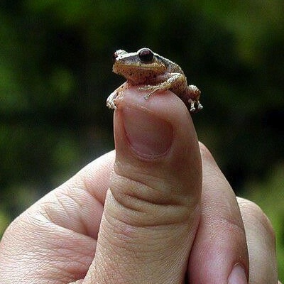
To understand if this factor was correlated with the presence of cell death in limbs, researchers looked for an amphibian naturally exposed to more oxygen than aquatic tadpoles. They found the Puerto Rican coqui frog, in a lab colony in the Museum of Comparative Zoology at Harvard University. Coqui frogs grow in terrestrial eggs without a tadpole stage.
Through a process called direct-development, coquis breathe oxygen from the air. Interestingly, they have dying cells in the interdigital region and a high production of ROS, just like chickens and mice. Data collected on them revealed their ecological features - such as where their embryos grow (in eggs) and how much oxygen surrounds each - has a direct effect on the presence of cell death in their limbs during their development.
The first step of a new evolutionary mechanism
However, other added steps might have taken place during evolution to make cell death a necessary mechanism in shaping hands and feet. "It is not clear if the dying cells are integrated to limb development in these frogs" said Mikiko Tanaka. "What we can say is that, during evolution, removal of the interdigital region by cell death became essential to shape the limbs of amniotes."
Researchers suggest that cell death in the limbs might have appeared as a byproduct of the high oxygen levels and ROS in this tissue, and were possibly a first step in this evolutionary process. They point out that further studies are necessary to investigate how cell death became an integral part of the limb development of amniotes, including modulation of pathways that respond to ROS and oxidative stress.
Highlights
• Interdigital cell death (ICD) is correlated with life-history strategy in tetrapods
• Atmospheric oxygen modulates local oxygen tension in the interdigital region
• High oxygen levels can induce ICD in an amphibian that typically lacks it
• Blood vessel density and Bmp signaling are critical for the appearance of ICD
Summary
Amphibians form fingers without webbing by differential growth between digital and interdigital regions. Amniotes, however, employ interdigital cell death (ICD), an additional mechanism that contributes to a greater variation of limb shapes. Here, we investigate the role of environmental oxygen in the evolution of ICD in tetrapods. While cell death is restricted to the limb margin in amphibians with aquatic tadpoles, Eleutherodactylus coqui, a frog with terrestrial-direct-developing eggs, has cell death in the interdigital region. Chicken requires sufficient oxygen and reactive oxygen species to induce cell death, with the oxygen tension profile itself being distinct between the limbs of chicken and Xenopus laevis frogs. Notably, increasing blood vessel density in X. laevis limbs, as well as incubating tadpoles under high oxygen levels, induces ICD. We propose that the oxygen available to terrestrial eggs was an ecological feature crucial for the evolution of ICD, made possible by conserved autopod-patterning mechanisms.
Authors
Ingrid Rosenburg Cordeiro, Kaori Kabashima, Haruki Ochi, Keijiro Munakata, Chika, Nishimori, Mara Laslo, James Hanken and Mikiko Tanaka.
Acknowlegements
The authors thank Dr. Kimberly Cooper, Dr. Shigeru Kuratani, and Dr. Atsushi Kawakami for critical comments; Dr. Akihiro Watanabe for donating newt larvae; Dr. Makoto Suzuki, Dr. Bau-Lin Huang, Dr. Akira Satoh, Dr. Takashi Kato, and Dr. Daisuke Sakai for technical advice; Dr. Cameron J. Koch for providing EF5 reagent and ELK3 antibodies; Mr. Matthew Gage for technical support; the Harvard Center for Biological Imaging and Dr. Douglas Richardson for imaging support; and the Biotechnology Center of Tokyo Institute of Technology for sequencing services. This work was supported by a Grant-in-Aid for Scientific Research (B) (16H04828), a Grant-in-Aid for Scientific Research on Innovative Areas (18H04818), the Takeda Science Foundation, and the Fujiwara Natural History Foundation to M.T.; Grant-in-Aid for Scientific Research (C) (16K07362), (B) (17KT0049), the Takeda Science Foundation, and the Suzuken Memorial Foundation to H.O.; and the U.S. National Science Foundation number DEB-1701591 to J.H.; the transgenic vector (ISXexEGFP) was provided by the Institute for Amphibian Biology (Hiroshima University, Japan) with support in part by the National Bio-Resource Project of the Japan Agency for Medical Research and Development (AMED) under grant number JP18km0210085.
Return to top of page.
| |
|
Jun 27 2019 Fetal Timeline Maternal Timeline News
 The Puerto Rican Coqui frog lays eggs and doesn't have a tadpole stage. Exposure to lots of oxygen during development affects how their limbs and digits form. CREDIT Pinterest.
|


