|
|
Developmental Biology - Brain Neurons
Brain Formation
The protein N-cadherin, may determine the location of neurons collecting into nerve bundles in our brain...
The human brain consists of countless neurons arranged into microscopic columns. But, the exact way this columnar structure organizes itself is elusive to neuroscientists. However, a research team led by Makoto Sato, has recently reported in the Journal of Neurosicence, the role of at least one specific protein.
The cortex, seat of most of our cerebral functions, forms the largest part of our brain and is divided into uncountable micro-columns of neurons.
The team, including Kanazawa University, Ryukoku University, Tokyo Institute of Technology and Imperial College London, did their research on Drosophila melanogaster (the fruit fly), whose brain visual centre structurally resembles the columnar arrangement of our own brain.
In order to visualize these columns of neurons, N-cadherin (Ncad) - a protein specific to the fly nervous system - was used to map spatial locations in the visual centre of the larval fly brain. The larva's visual centre is a donut-like structure which, as the fly matures into a pupa, begins stacking one donut shape on top of another transforming into a three-dimensional column.
Researchers carefully analysed these neuron columns to find a preponderance of three neuron types: R7, R8 and Mi1. While R7 was concentrated towards the center core of a column, R8 and Mi1 were arranged towards its outer edge. As the structure of the column was now clearer, what was left to understand was how column formation continues. Ncad was suspected to play a role as it is heavily present in all of these brain neurons.
Ncad was measured in all three neuron types, with more Ncad found in R7 neurons than peripheral neurons R8 and Mi1. As Ncad is known to give cells adhesive properties, the team concluded the levels of Ncad determined the location of each neuron type within a column with heavily adhesive neuron R7 forming the core of each column. To further confirm the affect of Ncad on column arrangement, the scientists then either completely removed Ncad or greatly increased it in neuron columns.
As expected, manipulation of Ncad disturbed the columnar assembly and positions of neurons.
Without Ncad being present, R7 neurons no longer migrated to the core of a column. However, when Ncad was greatly increased, the R8 and Mi1 neurons extended into the core as well, possibly due to Ncad's strong adhesive properties. So this study reveals how an adhesive protein in neurons affects their arrangement into columnar microstructures in the brain, a discovery which can help neuroscientists monitor healthy brain development and uncover other molecules involved in brain formation.
"Ncad-dependant differential adhesion and inter-layer interaction may be the essential mechanism underlying the 3D organization of column formation that is evolutionarily conserved acrossspecies from the fly optic lobe to mammalian brains," conclude the researchers.
Significance Statement
The columnar structure is a basic unit of the brain, but its developmental mechanism remains unknown. The medulla, the largest ganglion of the fly visual center, provides a unique opportunity to reveal the mechanisms of three-dimensional organization of the columns. We reveal that column formation is initiated by three core neurons that establish distinct concentric domains within a column. We demonstrate the in vivo evidence of N-cadherin-dependent differential adhesion among the core columnar neurons within a column along a two-dimensional layer in the larval medulla. The two-dimensional larval columns evolve to form three distinct layers in the pupal medulla. We propose the presence of mutual interactions among the three layers during formation of the three-dimensional structures of the medulla columns.
Abstract
Columnar structure is a basic unit of the brain, but the mechanism underlying its development remains largely unknown. The medulla, the largest ganglion of the Drosophila melanogaster visual center, provides a unique opportunity to reveal the mechanisms of three-dimensional organization of the columns. In this study, using N-cadherin (Ncad) as a marker, we reveal the donut-like columnar structures along the two-dimensional layer in the larval medulla that evolves to form three distinct layers in pupal development. Column formation is initiated by three core neurons, R8, R7, and Mi1, which establish distinct concentric domains within a column. We demonstrate that Ncad-dependent relative adhesiveness of the core columnar neurons regulates their relative location within a column along a two-dimensional layer in the larval medulla according to the differential adhesion hypothesis. We also propose the presence of mutual interactions among the three layers during formation of the three-dimensional structures of the medulla columns.
Authors
Olena Trush, Chuyan Liu, Xujun Han, Yasuhiro Nakai, Rie Takayama, Hideki Murakawa, Jose A. Carrillo, Hiroki Takechi, Satoko Hakeda-Suzuki, Takashi Suzuki and Makoto Sato.
Acknowlegements
The authors thank Hugo Bellen, Takashi Hayashi, Aljosha Nern, Takumi Suzuki and Tetsuo Yasugi for critical comments on the manuscript. We are grateful to Martin Heisenberg, Graeme Mardon, Hideji Murakoshi, Tadashi Uemura and S. Lawrence Zipursky for reagents and fly strains. We thank Bloomington Stock Center, Vienna Drosophila RNAi Center and DGRC, Kyoto for fly strains and Developmental Study Hybridoma Bank (DSHB) for antibodies. This work was supported by Grant-in-Aid for JSPS Research Fellow (to O.T.), CREST from JST, Grant-in-Aid for Scientific Research (B) from MEXT, Takeda Science Foundation and Cooperative Research of 'Network Joint Research Center for Materials and Devices' (to M.S.), and by Grant-in-Aid for Scientific Research on Innovative Areas from MEXT (To M.S. and T.S.).
Return to top of page.
| |
|
Jun 28 2019 Fetal Timeline Maternal Timeline News
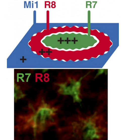 (TOP) Ncad R7, R8 and Mi1 form the basic structure of brain columns. The relative adhesiveness and Ncad expression level are in the order of R7 > R8 > Mi1. (BOTTOM) Visualization of terminal ends of R7 (GREEN) and R8 (RED) in the developing fly brain. CREDIT Kanazawa University
|


