|
|
Developmental Biology - 3D vision
3D Vision Brain Cells
Neurons found in insect brains that compute 3D distance and direction...
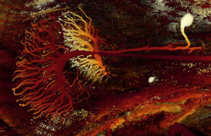
For the first time, stunning 3D images are captured of neurons in praying mantises. The work is published in Nature Communications.
In a specially-designed insect cinema, praying mantises were fitted with 3D glasses and shown 3D moving images of insect prey while their own brain activity was monitored. When an images of an insect came into striking range for a predatory attack by the mantis, scientist Ronny Rosner was able to record activity of individual neurons in the mantis brain.
"This helps us answer how insects achieve surprisingly complex behaviour with such tiny brains. Understanding their focusing ability helps us develop algorithms for robot and machine vision."
Ronny Rosner PhD, Institute of Neuroscience, Henry Wellcome Building for Neuroecology, Newcastle University, Framlington Place, Newcastle Upon Tyne, UK.
Stereopsis
Praying mantises use 3D perception, known as stereopsis, for hunting. Calculating the spatial difference between their two retinas, the mantis is able to compute distance and trigger a strike point when prey is within reach. Mantis neurons that record visual activity are photographed, extracted and stained, revealing their shape and allowed the team to identify four classes of neurons likely involved in mantis stereopsis.
"Despite their tiny size, mantis brains contain a surprising number of neurons that seem specialised for 3D vision. This suggests mantis depth perception is more complex than previously thought. As their brains are so much smaller than our own, we hope mantises can help us develop simpler algorithms for machine vision," explains Rosner.
"The properties within mantis brains are similar to what we see in the visual cortex of primates. When we see two very different species independently having evolved similar solutions, we know this must be a really good way of solving 3D vision.
We also found feedback loops within their 3D vision not seen in vertebrates. Our 3D vision may well include similar feedback loops — but are much easier to identify in less complex insect brains."
Jenny C. A. Read PhD, Institute of Neuroscience, Henry Wellcome Building for Neuroecology, Newcastle University, Framlington Place, Newcastle Upon Tyne, UK and project leader.
Abstract
A puzzle for neuroscience—and robotics—is how insects achieve surprisingly complex behaviours with such tiny brains. One example is depth perception via binocular stereopsis in the praying mantis, a predatory insect. Praying mantids use stereopsis, the computation of distances from disparities between the two retinal images, to trigger a raptorial strike of their forelegs when prey is within reach. The neuronal basis of this ability is entirely unknown. Here we show the first evidence that individual neurons in the praying mantis brain are tuned to specific disparities and eccentricities, and thus locations in 3D-space. Like disparity-tuned cortical cells in vertebrates, the responses of these mantis neurons are consistent with linear summation of binocular inputs followed by an output nonlinearity. Our study not only proves the existence of disparity sensitive neurons in an insect brain, it also reveals feedback connections hitherto undiscovered in any animal species.
Authors
Ronny Rosner, Joss von Hadeln, Ghaith Tarawneh and Jenny C. A. Read.
Acknowlegements
This work was funded by a Research Leadership Award RL-2012-019 from the Leverhulme Trust to J.R. We thank Dr Sasha Gartside and Dr Richard McQuade for providing equipment and advice when setting up the laboratory, Prof Uwe Homberg, Dr Claire Rind, Dr Peter Simmons, Prof Yoshifumi Yamawaki, Dr Sid Henriksen, Prof Keram Pfeiffer, Prof Ignacio Serrano-Pedraza and Prof David O’Carroll for helpful discussions, Prof Geraldine Wright for lending a micromanipulator, Dr Alex Laude and Dr Rolando Palmini from the Newcastle University Bioimaging Unit for help with the confocal microscopy, Katharina Wüst for providing praying mantids, Prof Ignacio Serrano-Pedraza for providing spectral filters, and Dr Bruce Cumming, Dr Vivek Nityananda, and Prof Geraldine Wright for helpful comments on the manuscript.
Return to top of page.
| |
|
Jul 2 2019 Fetal Timeline Maternal Timeline News
 A praying mantis visual neuron captured in 3D under microscope. CREDIT Newcastle University, UK.
|


