Welcome to The Visible Embryo
  o
 
The Visible Embryo Birth Spiral Navigation
   
Google  
Fetal Timeline--- -Maternal Timeline-----News-----Prescription Drugs in Pregnancy---- Pregnancy Calculator----Female Reproductive System

   
WHO International Clinical Trials Registry Platform

The World Health Organization (WHO) has a Web site to help researchers, doctors and patients obtain information on clinical trials.

Now you can search all such registers to identify clinical trial research around the world!


Home

History

Bibliography

Pregnancy Timeline

Prescription Drug Effects on Pregnancy

Pregnancy Calculator

Female Reproductive System

News

Disclaimer: The Visible Embryo web site is provided for your general information only. The information contained on this site should not be treated as a substitute for medical, legal or other professional advice. Neither is The Visible Embryo responsible or liable for the contents of any websites of third parties which are listed on this site.


Content protected under a Creative Commons License.
No dirivative works may be made or used for commercial purposes.


Pregnancy Timeline by SemestersDevelopmental TimelineFertilizationFirst TrimesterSecond TrimesterThird TrimesterFirst Thin Layer of Skin AppearsEnd of Embryonic PeriodEnd of Embryonic PeriodFemale Reproductive SystemBeginning Cerebral HemispheresA Four Chambered HeartFirst Detectable Brain WavesThe Appearance of SomitesBasic Brain Structure in PlaceHeartbeat can be detectedHeartbeat can be detectedFinger and toe prints appearFinger and toe prints appearFetal sexual organs visibleBrown fat surrounds lymphatic systemBone marrow starts making blood cellsBone marrow starts making blood cellsInner Ear Bones HardenSensory brain waves begin to activateSensory brain waves begin to activateFetal liver is producing blood cellsBrain convolutions beginBrain convolutions beginImmune system beginningWhite fat begins to be madeHead may position into pelvisWhite fat begins to be madePeriod of rapid brain growthFull TermHead may position into pelvisImmune system beginningLungs begin to produce surfactant
CLICK ON weeks 0 - 40 and follow along every 2 weeks of fetal development


Developmental Biology - Mitochondrial Disease

ANT Protein Mutations Fail to Supress Mitochondria

Mutations in ANT protein impair its ability to eliminate damaged mitochondria...


Mutations in the gene that encodes the protein adenine nucleotide translocator or ANT, jeopardize mitochondrial integrity by failing to eliminate damaged mitochondria. However, ANT also boosts the ability of mitochondria to produce chemical energy by increasing adenosine triphosphate (ATP) the fuel essential to all cells. These two contradictory behaviors are now understood to be part of ANT's 'quality control mechanism' over mitochondria.

New research from the University of Pennsylvania Medicine (Penn Medicine) has identified ANT as critical to mitochondrial quality control through the removal of damaged mitochondria — mitophagy — in addition to its ability to increase ATP. This is a newly defined behavior for the ANT protein.
Although ANT gene mutations cause disease, the research shows these mutations do not impact ANT's ability to produce ATP.

These findings are published in Nature Research, revealing this surprising new function of ANT, an otherwise very well-known protein.
"Our findings change the way we think about these disease-causing mutations and enable us to focus our attention on the right pathway. Now that we know these diseases are caused by defective quality control rather than a lack of ATP generation, we can start thinking about therapeutic approaches that improve quality control."

Zoltan Arany MD PhD, Professor of Cardiovascular Medicine, Perelman School of Medicine, University of Pennsylvania and corresponding author.

• Mitochondria are known as the powerhouse of the cell and generate most of the ATP needed to fuel cells. They play an important role in high-energy demanding organs, such as the heart and liver.

• Mitophagy occurs in defective mitochondria after damage or stress. ANT's role in mitophagy was not known until the Penn Medicine research.

• To understand all the components involved in mitophagy, researchers conducted a CRISPR/Cas9 genome-wide screening, using multiple reporter systems and pro-mitophagy triggers.
To their surprise, they found ANT is required for the quality control pathway of mitophagy in a number of cell types. In confirming follow-up tests, researchers found ANT promotes mitophagy independent of its role in producing ATP. This shows that the protein has two distinct jobs.

Upon this revelation, researchers designed tests to investigate whether mutations in the genes of ANT affected mitophagy.

• They find that disease-causing mutations in ANT cancel its ability to bind with protein complexes required to relocate the inner-membrane into the mitochondrial matrix, ultimately inhibiting mitophagy.

• Further studies showed that genetic removal of ANT suppresses mitophagy.

• Researchers also found mice genetically lacking ANT had blunted mitophagy and experienced a profound accumulation of atypical mitochondria.
"We were extremely surprised to find ANT participates in mitophagy, revealing it has two separate functions — ATP generation and quality control. While there's no magic bullet for improving its quality control process, we hope our findings lay the groundwork for future efforts examining how to offset suppression mitophagy and ultimately, prevent these mutations from causing disease."

Zoltan Arany MD PhD

Abstract
Mitochondrial homeostasis vitally depends on mitophagy, the programmed degradation of mitochondria. The roster of proteins known to participate in mitophagy remains small. We devised here a multidimensional CRISPR/Cas9 genetic screen, using multiple mitophagy reporter systems and pro-mitophagy triggers, and uncover numerous new components of Parkin-dependent mitophagy1. Unexpectedly, we identify the adenine nucleotide translocator (ANT) complex as required for mitophagy in multiple cell types. While pharmacological inhibition of ANT-mediated ADP/ATP exchange promotes mitophagy, genetic ablation of ANT paradoxically suppresses mitophagy. Importantly, ANT promotes mitophagy independently of its nucleotide translocase catalytic activity. Instead, the ANT complex is required for inhibition of the presequence translocase TIM23, leading to PINK1 stabilization, in response to bioenergetic collapse. ANT modulates TIM23 indirectly via interaction with TIM44, known to regulate peptide import through TIM232. Mice lacking ANT1 reveal blunted mitophagy and consequent profound accumulation of aberrant mitochondria. Disease-causing human mutations in ANT1 abrogate binding to TIM44 and TIM23 and inhibit mitophagy. Together, these data identify a novel and essential function for ANT as a fundamental mediator of mitophagy in health and disease.

Authors
Atsushi Hoshino, Wei-jia Wang, Shogo Wada, Chris McDermott-Roe, Chantell S. Evans, Bridget Gosis, Michael P. Morley, Komal S. Rathi, Jian Li, Kristina Li, Steven Yang, Meagen J. McMannus, Caitlyn Bowman, Prasanth Potluri, Michael Levin, Scott Damrauer, Douglas C. Wallace, Erika L. F. Holzbaur and Zoltan Arany.


Acknowledgments
This research was supported by HHMI, Project ALS, HSCI, Target ALS and the NINDS grant NIH5R01NS089742 to K.E. J.R.K. is the Project ALS Tom Kirchhoff Family Postdoctoral Fellow. B.N.D.-D. was supported by the Milton Safenowitz postdoctoral fellowship from the ALS Association. A. Burberry was supported by the US National Institutes of Health (1K99AG057808–01A1). D.A.M. was funded by the MGH training grant (5T32CA009216) and is grateful for the assistance of the Massachusetts ADRC neuropathology core in preparing tissue samples. B.J.W. is a New York Stem Cell Foundation – Robertson Investigator. We thank D. Cleveland for the generous gift of TDP-43 (FL9) antibody.

The work was supported, in part, by grants from the National Institutes of Health (HL094499, R37 NS060698, (NS021328, OD010944, MH108592, MH110185 and DK107667), U.S. Department of Defense (W81XWH-16-1-0401) and the established investigator award from the American Heart Association.

Penn Medicine is one of the world's leading academic medical centers, dedicated to the related missions of medical education, biomedical research, and excellence in patient care. Penn Medicine consists of the Raymond and Ruth Perelman School of Medicine at the University of Pennsylvania (founded in 1765 as the nation's first medical school) and the University of Pennsylvania Health System, which together form a $7.8 billion enterprise.

The Perelman School of Medicine has been ranked among the top medical schools in the United States for more than 20 years, according to U.S. News & World Report's survey of research-oriented medical schools. The School is consistently among the nation's top recipients of funding from the National Institutes of Health, with $425 million awarded in the 2018 fiscal year.

The authors thank the staff of the Penn Medicine Biobank including J. Weaver, D. Birtwell, H. Williams, P. Baumann, and M. Risman, as well as the Regeneron Genetics Center (RGC). The Penn Medicine Biobank was funded by a gift from the Smilow family and by the Penn Cardiovascular Institute and the Perelman School of Medicine. A.H. was supported by the Uehara Memorial Foundation, Mochida Memorial Foundation for Medical and Pharmaceutical Research and JSPS Overseas Research Fellowships. S.W. was supported by a fellowship from the ADA (1-16-PDF-117) and Toyobo Biotechnology Foundation. C.S.E. was supported by a Hanna H. Gray Fellowship from the Howard Hughes Medical Institute. E.L.F.H. was supported by the NIH NINDS (R37 NS060698). D.C.W. was supported by the NIH (NS021328, OD010944, MH108592, MH110185) and DOD (W81XWH-16-1-0401) Z.A. was supported by the NIH (HL094499, DK107667) and the AHA (Established Investigator Award).


Return to top of page.

Oct 24 2019   Fetal Timeline   Maternal Timeline   News  

Breast Cancer Awareness Month

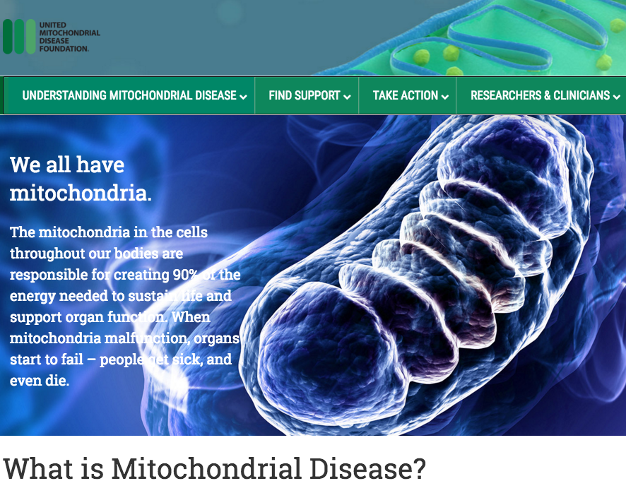
Mitochondria are in cells throughout our bodies. They are responsible for 90% of the energy needed to sustain life and support organ function. When mitochondria malfunction, organs start to fail.
CREDIT United Mitochondrial Diesase Foundation.


Phospholid by Wikipedia