|
|
Developmental Biology - Cell Fate
'Sleeping Cells' Can Be Harmful
New research into the mechanics of cell 'sleep & shutdown' may shed light on our own aging - and intervention...
A University of Arizona-led research team challenged the traditional understanding of cellular sleep to discover new information that might lead to interventions in our aging process.
As we age, more and more of our cells enter a coma-like state we call senescence. They can no longer divide to create new cells. Accumulation of senescent cells impairs normal tissue function and promotes aging. In contrast, many other cells in our body exist in a sleep-like state, called quiescence. These cells can be woken up or stimulated to divide in response to a trigger like a wound for example reversing quiescence and critical to repairing tissue as well as cell stability.
The research team found, contrary to traditional understanding, that as cells fall into deep sleep - they risk slipping into a complete shutdown.
According to paper co-author Guang Yao, an associate professor in the Department of Molecular and Cellular Biology, leader of the lab that produced the research: "Before, people thought cells slept to protect themselves from going into a coma-like state, thinking these two things are opposite. But we have demonstrated that sleep has different levels, and if it goes too deep, it will eventually go into a coma-like state of shutdown."
Researchers also discovered that patterns of gene expression can signal just how deeply a cell is sleeping. Learning to manipulate the depth of cellular sleep could lead to aging interventions.
"If you understand the mechanics of aging, you might be able to reverse or slow it," said Kotaro Fujimaki, doctoral student and first author on the paper published in the peer-reviewed journal Proceedings of the National Academy of Sciences of the United States of America, or PNAS.
Researchers gradually pushed cells into sleep of increasing depth using pharmaceuticals. Gene expression patterns revealed that as cell sleep deepened, its ability to break down and recycle cellular material, called the lysosomal-autophagy function, also fell. Consequently, cells underwent harmful chemical instability and stress, which can eventually cause a coma-like state.
When researchers cranked up this cellular recycling function, they observed reduced cell stress. The cells progressively moved into a shallow sleep, making them easier to reawaken and divide.
"By changing this recycler function, we can modulate the depth of cellular sleep."
Kotaro Fujimaki PhD, Department of Molecular and Cellular Biology, University of Arizona, Tucson, Arizona, USA;.
By analyzing how gene expression patterns change as cells go into deeper sleep, the team created a predictive model able to assign cells sleep depth scores between 1 to 10, to be applied to various cell types. Using this model, they also identified cells in a coma-like state undergo aging. This suggests that cells in deep sleep share similar gene expression features as those shutdown and aging.
Like a dimmer switch, cellular sleep ultimately exists on a spectrum of cell function between on-state (division) and off-state (shutdown), in a new paradigm proposed by the team.
According to Yao: "The dimmer controls how difficult it is to wake the cell back up for tissue repair and regeneration."
Researchers believe revealing the dimmer switch connecting deep cell sleep to coma-like shutdown, is the foundation for developing novel strategies, such as sliding the dimmer up, to slow aging down.
Significance
Reactivating sleep-like quiescent cells in the body (most notably stem cells) to divide is fundamental to tissue homeostasis and regeneration. Like sleep having shallow and deep stages, quiescence exhibits graded depths with different speeds and rates responding to stimulation signals. Understanding how quiescence depth is regulated is fundamental to our understanding of tissue repair and regeneration. Here we show that quiescence depth is regulated like a dimmer switch by lysosomes (cell organelles that break down and recycle biomolecules), through the lysosomal function of reducing oxidative stress. We developed a gene signature to predict quiescence depth and show that quiescence deepening likely represents a transition path from cell division to irreversibly arrested senescence, related to the aging process.
Abstract
The reactivation of quiescent cells to proliferate is fundamental to tissue repair and homeostasis in the body. Often referred to as the G0 state, quiescence is, however, not a uniform state but with graded depth. Shallow quiescent cells exhibit a higher tendency to revert to proliferation than deep quiescent cells, while deep quiescent cells are still fully reversible under physiological conditions, distinct from senescent cells. Cellular mechanisms underlying the control of quiescence depth and the connection between quiescence and senescence are poorly characterized, representing a missing link in our understanding of tissue homeostasis and regeneration. Here we measured transcriptome changes as rat embryonic fibroblasts moved from shallow to deep quiescence over time in the absence of growth signals. We found that lysosomal gene expression was significantly up-regulated in deep quiescence, and partially compensated for gradually reduced autophagy flux. Reducing lysosomal function drove cells progressively deeper into quiescence and eventually into a senescence-like irreversibly arrested state; increasing lysosomal function, by lowering oxidative stress, progressively pushed cells into shallower quiescence. That is, lysosomal function modulates graded quiescence depth between proliferation and senescence as a dimmer switch. Finally, we found that a gene-expression signature developed by comparing deep and shallow quiescence in fibroblasts can correctly classify a wide array of senescent and aging cell types in vitro and in vivo, suggesting that while quiescence is generally considered to protect cells from irreversible arrest of senescence, quiescence deepening likely represents a common transition path from cell proliferation to senescence, related to aging.
Authors
Kotaro Fujimaki, Ruoyan Li, Hengyu Chen, Kimiko Della Croce, Hao Helen Zhang, Jianhua Xing, Fan Bai, and Guang Yao.
Acknowledgments
Other University of Arizona co-authors were Kimiko Della Croce, an assistant staff scientist in the Department of Molecular and Cellular Biology, and Hao Helen Zhang, professor of mathematics. Also participating in the research were Ruoyan Li, Hengyu Chen and Bai Fan of Peking University, China; and Jianhua Xing of the University of Pittsburgh.
This work was supported by grants from the National Science Foundation, National Institutes of Health and the Chinese National Science and Technology Major Project and the Guangdong Province Key Research and Development Program.
Return to top of page.
| |
|
Nov 1 2019 Fetal Timeline Maternal Timeline News
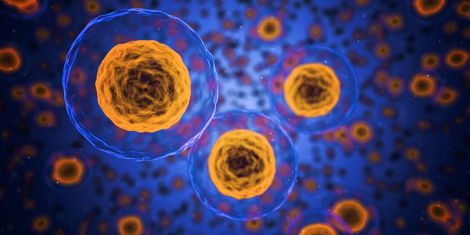
Like a dimmer switch, cell sleep ultimately controls cell function between 'on' (cell-division) and 'off' (cell-shutdown). Credit: Public Domain.
|


