|
|
Developmental Biology - Evolution of Single Cell to MultiCellular
When Single-Celled Microbes Became Complex
Animals evolved from single-celled ancestors, long before they diversifying into 30 or 40 distinct anatomical shapes...
When and how animals made the transition from single-celled microbes into complex multicellular organisms has been the focus of intense debate.
Until now, this question could only be addressed by studying living animals and their relatives. But, a research team has found evidence that a key step in this major evolutionary transition occurred long before complex animals appeared in the fossil record. They found fossilised embryos resembling multicellular stages in the life cycle of single-celled relatives of animals.
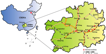
A team has discovered fossils named Caveasphaera in 609 million-year old rocks
in the Guizhou Province of South China.

Individual Caveasphaera fossils are only about half a millimeter in diameter, but X-ray microscopy revealed each was preserved all the way
down to their component cells.
"X-Ray tomographic microscopy works like a medical CT scanner, but allows us to see features that are less than a thousandth of a millimeter in size. We were able to sort the fossils into growth stages, reconstructing the embryology of Caveasphaera," says Kelly Vargas, from the University of Bristol's School of Earth Sciences.
"Our results show that Caveasphaera sorted its cells during embryo development, in just the same way as living animals do, including humans, but we have no evidence that these embryos developed into more complex organisms," adds Zongjun Yin, co-author from Nanjing Institute of Geology and Palaeontology in China.
"Caveasphaera had a life cycle like the close living relatives of animals, which alternate between single-celled and multicellular stages. However, Caveasphaera goes one step further, reorganising those cells during embryology. Caveasphaera is the earliest evidence of this most important step in the evolution of animals, which allowed them to develop distinct tissue layers and organs."
John Cunningham, University of Bristol and co-author.
Co-author Maoyan Zhu, also from Nanjing Institute of Geology and Palaeontology, is not totally convinced that Caveasphaera is an animal, adding another perspective: "Caveasphaera looks a lot like the embryos of some starfish and corals - we don't find the adult stages simply because they are harder to fossilise.
"This study shows amazing detail that can be preserved in the fossil record, but also the power of X-ray microscopes in uncovering secrets preserved in stone without destroying the fossils," adds Dr Federica Marone from the Paul Scherrer Institute in Switzerland.
"Caveasphaera shows features that look both like microbial relatives of animals and early embryo stages of primitive animals. We're still searching for more fossils that may help us to decide, added Professor Philip Donoghue, Co-author, also from the University of Bristol's School of Earth Sciences.
"Fossils of Caveasphaera tell us that animal-like embryonic development evolved long ago."
The discovery is published in Current Biology.
Highlights
• Caveasphaera is an enigmatic component of the 609-Ma Weng’an Biota of South China
• Yin et al. use X-ray tomography to characterize cellular structure and development
• Gastrulation-like cell division, ingression, detachment, and polar aggregation occur
• A holozoan affinity suggests the early evolution of metazoan-like development
Summary
The Ediacaran Weng’an Biota (Doushantuo Formation, 609 Ma old) is a rich microfossil assemblage that preserves biological structure to a subcellular level of fidelity and encompasses a range of developmental stages [1]. However, the animal embryo interpretation of the main components of the biota has been the subject of controversy [2, 3]. Here, we describe the development of Caveasphaera, which varies in morphology from lensoid to a hollow spheroidal cage [4] to a solid spheroid [5] but has largely evaded description and interpretation. Caveasphaera is demonstrably cellular and develops within an envelope by cell division and migration, first defining the spheroidal perimeter via anastomosing cell masses that thicken and ingress as strands of cells that detach and subsequently aggregate in a polar region. Concomitantly, the overall diameter increases as does the volume of the cell mass, but after an initial phase of reductive palinotomy, the volume of individual cells remains the same through development. The process of cell ingression, detachment, and polar aggregation is analogous to gastrulation; together with evidence of functional cell adhesion and development within an envelope, this is suggestive of a holozoan affinity. Parental investment in the embryonic development of Caveasphaera and co-occurring Tianzhushania and Spiralicellula, as well as delayed onset of later development, may reflect an adaptation to the heterogeneous nature of the early Ediacaran nearshore marine environments in which early animals evolved.
Authors
Zongjun Yin, Kelly Vargas, John Cunningham, Stefan Bengtson, Maoyan Zhu, Federica Marone, Philip Donoghue.
Acknowledgments
This research was funded through the Biosphere Evolution, Transitions and Resilience (BETR) program, which is co-funded by the Natural Environment Research Council (NERC) and Natural Science Foundation of China (NSFC).
Author Contributions
M.B.F. and F.C.-H. designed experiments. F.C.-H. performed experiments and data analysis.
Declaration of Interests
The authors declare no competing interest.
The research was supported by the National Institutes of Health (NIH F31EY028022-03, RO1EY019498, RO1EY013528, P30EY003176).
Return to top of page.
| |
|
Dec 2 2019 Fetal Timeline Maternal Timeline News
 These are computer models based on X-ray tomographic microscopy of the fossils, showing the successive stages of development. CREDIT Image by Franklin Caval-Holme, UC Berkeley
|


