|
|
Developmental Biology - Serotonin Reuptake Inhibitors (SSRIs)
Common Antidepressant Affects Baby
Early exposure to a common antidepressant (SSRI) during pregnancy changes sensory processing in neonatal mouse brain...
The serotonin (5HT) system and its proper development are critical to typical neurologic function, and implicated in a variety of neurodevelopmental processes and psychiatric conditions. 5HT modification is a common target for pharmaceuticals such as selective serotonin reuptake inhibitors (SSRIs), which are widely used for the treatment of depression and anxiety, including in an increasing proportion of pregnant women. Medical News Today.
• The scientific name for serotonin is 5-hydroxytryptamine, or 5-HT.
• It is mainly found in the brain, bowels, and blood platelets.
• Serotonin is used to transmit messages between nerve cells.
• It is thought to be active in constricting smooth muscles.
• It contributes to wellbeing and happiness, among other things, and as the precursor for melatonin, it helps regulate the body's sleep-wake cycles — our internal clock.
Exposure to antidepressants during pregnancy and the first weeks of life can alter that child's sensory processing well into adulthood. This new research in mice recently published December 2019 in eNeuro.
Physicians are increasingly prescribing a common antidepressant to their pregnant patients — even though the effect on the fetus isn't fully known.
A working theory of depression implicates the neurotransmitter serotonin because many depressed patients experience relief when prescribed SSRIs (selective serotonin reuptake inhibitors).
Whatever its role in depression, serotonin is critical for healthy brain development and function.
While previous research has shown changes in behavior and brain structure with prenatal and early life exposure to SSRIs, Rachel Rahn PhD, Departments of Radiology, Genetics and Psychiatry, Washington University School of Medicine, St. Louis, Missouri, with her team explored changes in brain activity.
After exposing mice to the SSRI fluoxetine during gestation and the first two weeks following birth, the team used optical imaging to examine those mouse brains exposed to fluoxetine as compared to control (untreated/normal) mice.
In a resting state, the brains of both sets of mice were nearly identical. However, when their front paws were electrically stimulated, the fluoxetine-exposed mice displayed abnormal brain activity in sensory areas.
The effect was observed in adult mice, suggesting their developmental exposure to SSRIs while in utero caused long-term changes to sensory processing.
Abstract
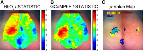
Epidemiological studies have found an increased incidence of neurodevelopmental disorders in populations prenatally exposed to selective serotonin reuptake inhibitors (SSRIs). Optical imaging provides a minimally invasive way to determine if perinatal SSRI exposure has long-term effects on cortical function. Herein we probed the functional neuroimaging effects of perinatal SSRI exposure in a fluoxetine (FLX)-exposed mouse model. While resting-state homotopic contralateral functional connectivity was unperturbed, the evoked cortical response to forepaw stimulation was altered in FLX mice. The stimulated cortex showed decreased activity for FLX versus controls, by both hemodynamic responses [oxyhemoglobin (HbO2)] and neuronal calcium responses (Thy1-GCaMP6f fluorescence). Significant alterations in both cortical HbO2 and calcium response amplitude were seen in the cortex ipsilateral to the stimulated paw in FLX as compared to controls. The cortical regions of largest difference in activation between FLX and controls also were consistent between HbO2 and calcium contrasts at the end of stimulation. Taken together, these results suggest a global loss of response signal amplitude in FLX versus controls. These findings indicate that perinatal SSRI exposure has long-term consequences on somatosensory cortical responses.
Significance Statement
Use of selective serotonin reuptake inhibitors (SSRIs) by pregnant women has increased in the past decades, raising questions regarding the long-term effects of SSRI usage during pregnancy on offspring. In order to isolate the effect of perinatal SSRI exposure from genetic variables or a maternal psychiatric diagnosis, we used in vivo functional neuroimaging to examine adult cortical function both at rest and in response to a somatosensory input in a mouse model of perinatal SSRI exposure. Our mouse model displayed no global disruption of brain function at rest when compared to controls, while its cortical response to a somatosensory stimulus was reduced, as measured by both hemodynamics and excitatory calcium signaling.
Authors
Rachel M. Rahn, Susan E. Maloney, Lindsey M. Brier, Joseph D. Dougherty and Joseph P. Culver
Return to top of page.
| |
|
Jan 2 2020 Fetal Timeline Maternal Timeline News
 Pictured are the outcomes of HbO2 and calcium response on mouse brains after electrical stimulation to left paw. Results are superimposed over brain scans. A: HbO2 t statistical map of FLX-VEH {fluoxetine (Flx), one of the most prescribed SSRIs. B: GCaMP6f t {which enables ratiometric imaging of Ca2+} statistical map of FLX-VEH C: Normal brain reflecting HbO2 and GCaMP6f (BLUE is HbO2; YELLOW is GCaMP6f; GREEN is both).
|


