Welcome to The Visible Embryo
  o
 
The Visible Embryo Birth Spiral Navigation
   
Google  
Fetal Timeline--- -Maternal Timeline-----News-----Prescription Drugs in Pregnancy---- Pregnancy Calculator----Female Reproductive System

   
WHO International Clinical Trials Registry Platform

The World Health Organization (WHO) has a Web site to help researchers, doctors and patients obtain information on clinical trials.

Now you can search all such registers to identify clinical trial research around the world!


Home

History

Bibliography

Pregnancy Timeline

Prescription Drug Effects on Pregnancy

Pregnancy Calculator

Female Reproductive System

News

Disclaimer: The Visible Embryo web site is provided for your general information only. The information contained on this site should not be treated as a substitute for medical, legal or other professional advice. Neither is The Visible Embryo responsible or liable for the contents of any websites of third parties which are listed on this site.


Content protected under a Creative Commons License.
No dirivative works may be made or used for commercial purposes.


Pregnancy Timeline by SemestersDevelopmental TimelineFertilizationFirst TrimesterSecond TrimesterThird TrimesterFirst Thin Layer of Skin AppearsEnd of Embryonic PeriodEnd of Embryonic PeriodFemale Reproductive SystemBeginning Cerebral HemispheresA Four Chambered HeartFirst Detectable Brain WavesThe Appearance of SomitesBasic Brain Structure in PlaceHeartbeat can be detectedHeartbeat can be detectedFinger and toe prints appearFinger and toe prints appearFetal sexual organs visibleBrown fat surrounds lymphatic systemBone marrow starts making blood cellsBone marrow starts making blood cellsInner Ear Bones HardenSensory brain waves begin to activateSensory brain waves begin to activateFetal liver is producing blood cellsBrain convolutions beginBrain convolutions beginImmune system beginningWhite fat begins to be madeHead may position into pelvisWhite fat begins to be madePeriod of rapid brain growthFull TermHead may position into pelvisImmune system beginningLungs begin to produce surfactant
CLICK ON weeks 0 - 40 and follow along every 2 weeks of fetal development


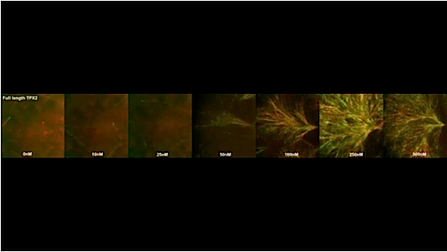
Developmental Biology - Cell Division

Princeton Recreates A Key Step In Cell Division

Recreating key cell division step that defines a protein elevated in cancer...


Research at Princeton University has successfully recreated a key process in cell division in the lab. They've been able to uncover the vital role played by a protein elevated in over 25% of all cancers. The findings are described in a pair of papers published in eLife and Nature Communications. These processes are key to creating the entire cell division machinery leading to new therapies to prevent the growth of cancer cells.
When cells divide, a spindle-like structure composed of thousands of filaments called microtubules attaches to chromosomes, pulling equal numbers of chromosomes into each newly forming cell.

Each microtubule is assembled from individual tubulin molecules and, because errors in chromosome segregation can lead to cancer, it is vital they assemble into microtubules at the right time and place to form a functional spindle apparatus.

• Branching microtubule nucleation, in which a new microtubule forms from the side of an existing tubule, is crucial to this process.

• This is because it allows the cell to form large numbers of microtubules that all point toward chromosomes, enabling their capture by the spindle.

Branching microtubule nucleation depends on several pieces of molecular machinery. One piece, called the gamma-tubulin ring complex (y-TuRC), initiates the assembly of tubulin molecules into microtubules. Another, known as the augmin complex, recruits y-TuRC to the side of existing microtubules.

A protein called TPX2, whose levels are elevated in over 25% of all cancers, is also involved in branching microtubule nucleation. Elevated TPX2 levels lead to both aberrant microtubule assembly in cells and poor outcomes in cancer patients. But, how TPX2 works with augmin and y-TuRC to mediate branching microtubule nucleation and spindle assembly has remained unknown.

TPX2 on microtubules film clip

TPX2 behaves like a liquid in promoting branching microtubule nucleation.
The protein TPX2 increases in concentration from left to right in this video clip.

"To better understand the mechanism of branching microtubule nucleation, we set out to reconstitute the process outside of the cell using purified proteins," explains Sabine Petry, assistant professor of molecular biology at Princeton.

In the eLife study, graduate students Raymundo Alfaro-Aco and Akanksha Thawani describe how they recreated branching microtubule nucleation in a test tube.
• One key finding from the study is that, like augmin, TPX2 can bind to microtubules and recruit y-TuRC to initiate branching microtubule nucleation.

• Another surprising finding was that TPX2 also helps recruit augmin to microtubules, further enhancing the recruitment of y-TuRC.

"Branching microtubule nucleation therefore occurs most efficiently when augmin, TPX2, and y-TuRC are all present. Surprisingly, TPX2 lies at the very heart of controlling this reaction, despite being only a single protein amongst the large multi-subunit complexes augmin and y-TuRC."


Raymundo Alfaro-Aco PhD, Department of Molecular Biology, Princeton University, Princeton, New Jersey, USA.

In the Nature Communications paper, Petry and her former graduate student Matthew King further reveal that TPX2 behaves like a liquid in promoting branching microtubule nucleation.
Specifically, TPX2 forms a liquid layer on the surface of existing microtubules that beads up into tubulin-containing droplets, much like morning dew on spider webs.

Researchers found that TPX2 and tubulin can condense together to form liquid-like droplets through a phase-separation mechanism identical to the one that causes oil droplets to form in water. New microtubules can form from these TPX2-tubulin droplets and, because the droplets condense on the surface of existing microtubules, this results in the formation of branched microtubule structures.
"The study suggests that the co-condensation of TPX2 and tubulin creates a local reservoir of tubulin on a pre-existing microtubule that may be necessary to efficiently promote branching microtubule nucleation."

Matthew R. King PhD, Department of Biomedical Engineering, Washington University, Saint Louis, Missouri; Department of Molecular Biology, Princeton University, Princeton, New Jersey, USA.

Together, both studies reveal that TPX2, somewhat overlooked before, is the lynchpin of branching microtubule nucleation. It travels to microtubules first to assemble all of the other components that ultimately give rise to a branching microtubule, and it does this while behaving like a liquid.

Petry and colleagues think that cellular signals may regulate TPX2 condensation to ensure that it only occurs when a cell is dividing and needs to form a spindle.
"Our findings on the behavior of TPX2 may guide future therapeutic efforts aimed at modulating cell division. In addition, our reconstitution of branching microtubule nucleation is an important step toward reconstituting the entire spindle apparatus, as well as other cellular structures that depend on this microtubule assembly pathway."

Sabine Petry PhD, Department of Molecular Biology, Princeton University, Princeton, New Jersey, USA.

Abstract Nature Communications
Phase separation of substrates and effectors is proposed to enhance biological reaction rates and efficiency. Targeting protein for Xklp2 (TPX2) is an effector of branching microtubule nucleation in spindles and functions with the substrate tubulin by an unknown mechanism. Here we show that TPX2 phase separates into a co-condensate with tubulin, which mediates microtubule nucleation in vitro and in isolated cytosol. TPX2-tubulin co-condensation preferentially occurs on pre-existing microtubules, the site of branching microtubule nucleation, at the endogenous and physiologically relevant concentration of TPX2. Truncation and chimera versions of TPX2 suggest that TPX2-tubulin co-condensation enhances the efficiency of TPX2-mediated branching microtubule nucleation. Finally, the known inhibitor of TPX2, the importin-a/ß heterodimer, regulates TPX2 condensation in vitro and, consequently, branching microtubule nucleation activity in isolated cytosol. Our study demonstrates how regulated phase separation can simultaneously enhance reaction efficiency and spatially coordinate microtubule nucleation, which may facilitate rapid and accurate spindle formation.

Authors
Matthew R. King and Sabine Petry.


Acknowledgments
The work published in both papers was supported by the National Institutes of Health (NIH), the Pew Scholars Program in the Biomedical Sciences, and the David and Lucile Packard Foundation. The work published in eLife was additionally supported by a Howard Hughes Medical Institute Gilliam Fellowship, a National Science Foundation Graduate Research Fellowship, and an American Heart Association predoctoral fellowship.

The authors thank members of the Petry Lab for helping with this work, including Ray Alfaro-Aco, Michael Rale, Mohammad Safari, Akanksha Thawani, and Sagar Setru. We are especially grateful to Cliff Brangwynne for experimental suggestions and the Petry Lab, Ibrahim Cisse, and Kassandra Ori-McKenney for feedback on the manuscript. This work was supported by a Ph.D. training grant T32GM007388 by NIGMS of the National Institutes of Health (to M.R.K.), as well as the New Innovator Award of NIGMS of the National Institutes of Health (DP2), the Pew Scholars Program in the Biomedical Sciences, and the David and Lucile Packard Foundation (all to S.P.).


Return to top of page.

Jan 22 2020   Fetal Timeline   Maternal Timeline   News 


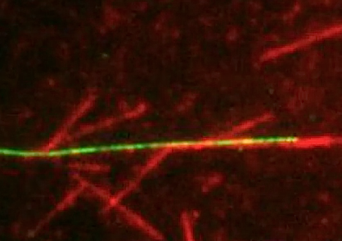
In the experiment, new microtubules formed when TPX2 condensed with tubulin into
liquid-like droplets. CREDIT Raymundo Alfaro-Aco, Sabine Petry, Princeton University


Phospholid by Wikipedia