|

CLICK ON weeks 0 - 40 and follow along every 2 weeks of fetal development
|
||||||||||||||||||||||||||||
|
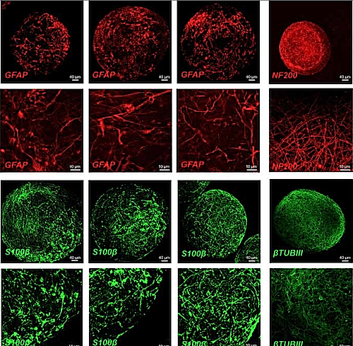
Developmental Biology - Preterm Birth Common Antidepressant Harms Lab 'Mini-Brains Hartung and colleagues developed mini-brains in order to model early human brain growth. These tiny clumps of brain tissue are made by taking cells from adult humans, often from their skin, and transforming them into stem cells, and then biochemically nudging the stem cells to develop into young brain cells. The mini-brains form a rudimentary brain-like organization over a period of a few months. Because they are made of human cells, they may be more likely to predict effects on the human brain — and because they can be mass-produced in the lab, they are much cheaper to work with than animals. A set of animal toxicology tests for a single chemical costs about $1.4 million on average, the authors note, which explains why the vast majority of chemicals used in drugs and other consumer products have never been tested for toxicity. In contrast, toxicity testing using mini-brains costs only a few thousand dollars. In the new study, the scientists used mini-brains to test for neurodevelopmental effects of paroxetine. It and other antidepressants in its class, known as SSRIs or selective serotonin reuptake inhibitors, are among the world's most commonly prescribed drugs, accounting for at least hundreds of millions of prescriptions annually. The research team exposed mini-brains to two different concentrations of paroxetine over eight weeks as BrainSphere clumps of tissue developed. Both concentrations were within the therapeutic range for blood levels of the drug in humans. In these experiments, researchers also used two different sets of mini-brains, each derived from a different stem cell. They found that while paroxetine didn't appear to have a significant neuron-killing effect, at higher concentrations it reduced levels of a protein called synaptophysin, which is a key component and marker of synapses — by up to 80%. These effects suggest the drug might hinder the normal formation of interconnections among developing neurons — a result that could conceivably underlie autism or other disorders. The study also shows the broader potential of mini-brains-based testing to detect adverse effects of drugs on the developing brain. Hartung: "In this report, we were able to show that testing with mini-brains can reveal relatively subtle neurodevelopmental effects, not just obvious effects, of a chemical. Whether Paroxetine causes autism has been a decade-long debate, which could not be settled with animal tests or epidemiological analyses. So we see mini-brains as technology for broader assessment of the risks of common drugs and chemicals, including those that might be contributing to the autism epidemic." Hartung and colleagues recently received a grant from the U.S. Environmental Protection Agency to develop their technology as an alternative to animal testing. Abstract Selective serotonin reuptake inhibitors (SSRIs) are frequently used to treat depression during pregnancy. Various concerns have been raised about the possible effects of these drugs on fetal development. Current developmental neurotoxicity (DNT) testing conducted in rodents is expensive, time-consuming, and does not necessarily represent human pathophysiology. A human, in vitro testing battery to cover key events of brain development, could potentially overcome these challenges. In this study, we assess the DNT of paroxetine—a widely used SSRI which has shown contradictory evidence regarding effects on human brain development using a versatile, organotypic human induced pluripotent stem cell (iPSC)-derived brain model (BrainSpheres). At therapeutic blood concentrations, which lie between 20 and 60 ng/ml, Paroxetine led to an 80% decrease in the expression of synaptic markers, a 60% decrease in neurite outgrowth and a 40–75% decrease in the overall oligodendrocyte cell population, compared to controls. These results were consistently shown in two different iPSC lines and indicate that relevant therapeutic concentrations of Paroxetine induce brain cell development abnormalities which could lead to adverse effects. Authors Xiali Zhong, Georgina Harris, Lena Smirnova, Valentin Zufferey, Rita de Cássia da Silveira e Sá, Fabiele Baldino Russo, Patricia Cristina Baleeiro Beltrao Braga, Megan Chesnut, Marie-Gabrielle Zurich, Helena Hogberg, Thomas Hartung and David Pamies. Acknowledgements The study was supported by funding from the European Union's Horizon 2020 research and innovation program (grant No. 487 681002). Co-authors Thomas Hartung, Helena Hogberg, and David Pamies are named inventors on a patent by Johns Hopkins University on the production of mini-brains, which is licensed to AxoSim, New Orleans, Louisiana. The co-authors consult for AxoSim and Thomas Hartung is a shareholder. David Pamies, who was corresponding author of the paper, worked at the Center for Alternatives to Animal Testing during the study and is now at the University of Lausanne. The study was supported by funding from the European Union's Horizon 2020 research and innovation program (grant No. 487 681002). Return to top of page. | Feb 21 2020 Fetal Timeline Maternal Timeline News  BrainSpheres treated at differentiation - 8 weeks -with 20 or 60 ng/ml Paroxetine. Represent (A) Percentage of viable cells in Paroxetine-treated BrainSpheres as measured by assays (B) Mitochondrial membrane potential measured (MMP) by mitotracker assay (C) Representative images of mitotracker assays (D) Immunohistochemistry with astrocyte markers (GFAP, S100ß) & neuronal markers (NF200, ßTUBIII). RED—PsigA-RFP and GREEN—Psig B-YFP. CREDIT the authors.
|
||||||||||||||||||||||||||||

