|
|
Developmental Biology - COVID-19
Harnessing the Power of Cord Blood
MicroRNA miR-125a switches cord blood into stem cells...
https://www.eurekalert.org/pub_releases/2016-07/uhn-scs071316.php
International stem cell scientists, co-led in Canada by Dr. John Dick and in the Netherlands by Dr. Gerald de Haan, have discovered the switch to harness the power of cord blood and potentially increase the supply of stem cells for cancer patients needing transplantation therapy to fight their disease.
Proof-of-concept findings, published online today in Cell Stem Cell provide a viable new approach to making more stem cells from cord blood, which is available through public cord blood banking, says co-principal investigator John Dick, Senior Scientist, Princess Margaret Cancer Centre, University Health Network (UHN). Dr. Dick is also Professor, Department of Molecular Genetics, University of Toronto, and holds a Canada Research Chair in Stem Cell Biology. The co-principal investigator was stem cell scientist Gerald de Haan, Scientific Co-Director, European Institute for the Biology of Ageing, University Medical Centre Groningen, the Netherlands. Dr. Dick talks about their joint research at https://youtu.be/cpEmKnjkb9s.
"Stem cells are rare in cord blood and often there are not enough present in a typical collection to be useful for human transplantation. The goal is to find ways to make more of them and enable more patients to make use of blood stem cell therapy," says Dr. Dick. "Our discovery shows a method that could be harnessed over the long term into a clinical therapy and we could take advantage of cord blood being collected in various public banks that are now growing across the country."
Currently, patients needing stem cell transplants are matched to an adult donor with a compatible immune system through international registry services. But worldwide, many thousands of patients are unable to get stem cell transplants needed to combat blood cancers such as leukemia because there is no donor match.
"About 40,000 people receive stem cell transplants each year, but that represents only about one-third of the patients who require this therapy," says Dr. Dick. "That's why there is a big push in research to explore cord blood as a source because it is readily available and increases the opportunity to find tissue matches. The key is to expand stem cells from cord blood to make many more samples available to meet this need. And we're making progress."
Although there is much research into expanding the rare stem cells present in cord blood, the Dick-de Haan teams took a different approach. When a stem cell divides it makes a lot of progenitor cells immediately downstream that retain key properties of being able to develop into every one of the 10 mature blood cell types, but they have lost the critical ability to self-renew (keeping on replenishing the stem cell pool) that all true stem cells possess.
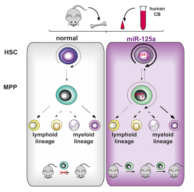
In the lab, analysing murine and human models of blood development, the teams discovered that microRNA (mirR-125a) is a genetic switch that is normally on in stem cells and controls self-renewal; this normally gets turned off in the progenitor cells.
"Our work shows that if we artificially throw the switch on in those downstream cells, we can endow them with stemness and they basically become stem cells and can be maintained over the long term," says Dr. Dick.
In 2011, Dr. Dick isolated a human blood stem cell in its purest form - as a single stem cell capable of regenerating the entire blood system, providing a more detailed road map of the human blood development system, and opening the door to capturing the power of these life-producing cells to treat cancer and other debilitating diseases more effectively.
Dr. Dick is also Senior Scientist at the McEwen Centre for Regenerative Medicine (UHN) and Director of the Cancer Stem Cell Program at the Ontario Institute for Cancer Research.
Stem cells were first discovered in Toronto in 1961 at the Princess Margaret by Drs. James Till and the late Ernest McCulloch - a discovery that launched a new field of science and formed the basis of all stem cell research that continues to this day.
Abstract Highlights
• Enforced expression of miR-125 increases hematopoietic stem frequency in vivo
• miR-125 induces stem cell potential in murine and human progenitor cells
• miR-125 represses, among others, targets of the MAP kinase signaling pathway
• miR-125 function and targets are conserved in human and mouse
Summary
Umbilical cord blood (CB) is a convenient and broadly used source of hematopoietic stem cells (HSCs) for allogeneic stem cell transplantation. However, limiting numbers of HSCs remain a major constraint for its clinical application. Although one feasible option would be to expand HSCs to improve therapeutic outcome, available protocols and the molecular mechanisms governing the self-renewal of HSCs are unclear. Here, we show that ectopic expression of a single microRNA (miRNA), miR-125a, in purified murine and human multipotent progenitors (MPPs) resulted in increased self-renewal and robust long-term multi-lineage repopulation in transplanted recipient mice. Using quantitative proteomics and western blot analysis, we identified a restricted set of miR-125a targets involved in conferring long-term repopulating capacity to MPPs in humans and mice. Our findings offer the innovative potential to use MPPs with enhanced self-renewal activity to augment limited sources of HSCs to improve clinical protocols.
Authors
Edyta E. Wojtowicz, Eric R. Lechman, Karin G. Hermans, Erwin M. Schoof, Erno Wienholds, Ruth Isserlin, Peter A. van Veelen, Mathilde J.C. Broekhuis, George M.C. Janssen, Aaron Trotman-Grant, Stephanie M. Dobson, Gabriela Krivdova, Jantje Elzinga, James Kennedy, Olga I. Gan, Ankit Sinha, Vladimir Ignatchenko, Thomas Kislinger, Bertien Dethmers-Ausema
Ellen Weersing, Mir Farshid Alemdehy, Hans W.J. de Looper, Gary D. Bader, Martha Ritsema, Stefan J. Erkeland, Leonid V. Bystrykh and John E. Dick.
Acknowledgements
The authors thank H. Moes, G. Mesander, and R. J. van der Lei for expert cell-sorting assistance; Klaas Sjollema from the UMCG Microscopy and Imaging Center (UMIC) for advice; and E. Verovskaya and R.P. van Os for discussions and assistance in the laboratory. The authors also thank the obstetrics units of Trillium Health Partners (Missisauga and Credit Valley sites) for the cord blood units and the UHN/Sick Kids and PMH Flow Cytometry Facilities for cell sorting. We would also like to thank Dr. K. Itoh (Kyoto University, Japan) for kindly providing MS-5 stromal cells.
This work was supported by a Rubicon fellowship from the Netherlands Organization for Scientific Research (to K.G.H.), a Canadian Institutes for Health Research (CIHR) fellowship (to K.G.H.), and grants from Netherlands Organization for Scientific Research (to G.d.H), EuroCSCTraining Eurocancer Stemcell Training Network ITN-FP7-Marie Curie Action 264361 (to E.E.W.), and the Netherlands Institute for Regenerative Medicine. E.M.S. is an EMBO Postdoctoral Fellow (ALTF 1595-2014) and is co-funded by the European Commission (LTFCOFUND2013, GA-2013-609409) and Marie Curie Actions. The work in J.E.D.’s lab is funded by the CIHR, Canadian Cancer Society Research Institute, Terry Fox Foundation, Genome Canada through the Ontario Genomics Institute, Ontario Institute for Cancer Research, a Canada Research Chair, the Princess Margaret Hospital Foundation, and the Ontario Ministry of Health and Long Term Care (OMOHLTC). The views expressed in this manuscript do not necessarily reflect those of the OMOHLTC.
The Canadian team's research published today was funded by the Canadian Institutes of Health Research, the Canadian Cancer Society Research Institute, the Terry Fox Foundation, the Ontario Institute for Cancer Research, the Canada Research Chair in Stem Cell Biology, The Princess Margaret Cancer Foundation, and the Ontario Ministry of Health and Long-term Care.
About Princess Margaret Cancer Centre, University Health Network
The Princess Margaret Cancer Centre has achieved an international reputation as a global leader in the fight against cancer and delivering personalized cancer medicine. The Princess Margaret, one of the top five international cancer research centres, is a member of the University Health Network, which also includes Toronto General Hospital, Toronto Western Hospital, Toronto Rehabilitation Institute and the Michener Institute for Education; all affiliated with the University of Toronto. For more information, go to http://www.theprincessmargaret.ca or http://www.uhn.ca.
Return to top of page.
| |
|
May 18 2020 Fetal Timeline Maternal Timeline News
 Umbilical cord blood cells with miR-125 lentiviral vector (miR-125OE) — or an empty control vector (n = 10) are injected into thefemur (upper leg bone) and distant bone marrow (BM) sites. RED and GRAY represent experiments using two distinct human Cord Bloods. Color-matched bars
represent input levels of mOrange+ cells at time of transplant in each experiment.
HSC = Human Stem Cell; MPP = Multipotent Progenitors cells CREDIT The Authors.
|


