|
|
Developmental Biology - Placental Pump Function
Contractions In Human Placenta Explained
New insight into blood pumping patterns of the placenta show how crucial it is to fetal development...
High-resolution imaging of the human placenta gives new insight into its control over crucial maternal and fetal blood circulation during development. The new study published May, 28 2020 in the open-access journal PLOS Biology, was written by Penny Gowland PhD, University of Nottingham, United Kingdom based on work with her colleagues.
Their findings improve our understanding of the function of this understudied organ, both in healthy pregnancies as well as in serious medical conditions — such as pre-eclampsia.
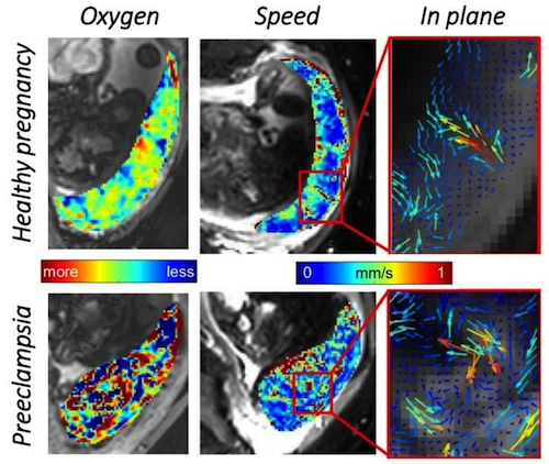
Using a technique called magnetic resonance imaging (MRI) to study fetal growth and development, Gowland and colleagues recorded the precise movement and oxygenation of blood in the human placenta. They observed relatively uniform and high oxygenation levels across the whole maternal contribution to the placenta, with a decrease in velocity of blood flow as it enters the placenta, reducing pressure to the fetus.
This finding suggests how efficiently oxygen is delivered from the mother to her fetus. Measuring how rapidly blood drains from veins in the placenta, crucial to proper circulation, was largely neglected by past studies.
Moreover, their discovery of a new physiological phenomenon, named the utero-placental pump, shows how the placenta and underlying uterine wall contract independent of the uterus.
Such strong contractions facilitate better blood flow through the placenta — and the fetus.
Finally, researchers also identified altered patterns of blood movement associated with pre-eclamptic pregnancies. Pre-eclampsia is characterized by high blood pressure and often a significant amount of protein in the mother's urine. Such conditions increase risk of poor outcomes for both mother and baby.
According to the new results, placental models will be improved - helping to optimize MRI protocols — and identify placental abnormalities earlier.
Abstract
We have used magnetic resonance imaging (MRI) to provide important new insights into the function of the human placenta in utero. We have measured slow net flow and high net oxygenation in the placenta in vivo, which are consistent with efficient delivery of oxygen from mother to fetus. Our experimental evidence substantiates previous hypotheses on the effects of spiral artery remodelling in utero and also indicates rapid venous drainage from the placenta, which is important because this outflow has been largely neglected in the past. Furthermore, beyond Braxton Hicks contractions, which involve the entire uterus, we have identified a new physiological phenomenon, the ‘utero-placental pump’, by which the placenta and underlying uterine wall contract independently of the rest of the uterus, expelling maternal blood from the intervillous space.
Authors
Neele S. Dellschaft, George Hutchinson, Simon Shah, Nia W. Jones, Chris Bradley, Lopa Leach, Craig Platt, Richard Bowtell and Penny A. Gowland.
Acknowledgements
Funding: This work was funded by the Human Placenta Project NIH grant 1U01HD087202-01 (PAG, NWJ, and RB).
The funders had no role in study design, data collection and analysis, decision to publish, or preparation of the manuscript.
Competing Interests: The authors have declared that no competing interests exist.
Return to top of page.
| |
|
Jun 1 2020 Fetal Timeline Maternal Timeline News
 These MRI screens show blood oxygen and speed of blood movement within the placenta for both a (TOP) healthy pregnancy and a (BOTTOM) preeclamptic pregnancy. - CREDIT Penny Gowland.
|


