|
|
Developmental Biology - Primary Cilia
'Antenna' Cover Neuron Cells
When these cell antenna are too few in number or too short in length, Fragile X syndrome and autism may occur. However, it may be a possible to increase their number...
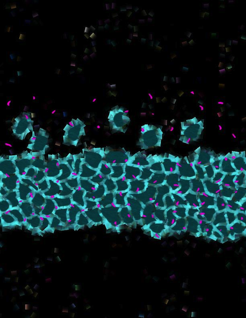
Structures called primary cilia act like antenna on the cell surface of neurons, detecting signals from other cells. They are also known to be critical to the maturation of neuronal cells in the DG or Dentate gyrus of the brain.
However, when there are fewer of them and/or they are of shorter than normal length, mice are born with Fragile X syndrome, according to research from the University of Texas Health Science Center, San Antonio (UT Health San Antonio). The study was just published July 30 in the journal Stem Cell Reports.
Fragile X syndrome is a genetic disorder often accompanied by mild to severe intellectual disability. Autism spectrum disorders also frequently occur in affected children. Better understanding of the affects of fewer primary cilia on Fragile X syndrome and autism may lead to new therapies for increasing their numbers. Possibly to even reversing these neurodevelopmental defects, suggests study senior author Hye Young Lee PhD.
The research focused on primary cilia located in a brain structure called the dentate gyrus. It is part of the hippocampus, a learning and memory command center. Dr. Lee and her colleagues found that reduction in primary cilia was specific to the dentate gyrus.
The dentate gyrus is one of two brain structures that contain neuronal stem cells — serving as a nursery for newly forming neurons. Neurons depend on primary cilia to receive and send signals enabling their role.
Previous to this study, primary cilia had not been linked to Fragile X syndrome.
"If we can confirm how primary cilia work in the newborn neuron, and how they contribute to Fragile X syndrome, our next step will be to promote their growth. There are drugs that can do this, and could be potential therapies for Fragile X syndrome and other neurodevelopmental disorders. Multiple studies are beginning to show that some neurodevelopmental disorders, such as autism, can be reversed or be made less severe with treatment."
Hye Young Lee PhD,
Department of Cellular and Integrative Physiology, University of Texas Health Science Center, San Antonio, Texas, USA.
Highlights
• Primary cilia are significantly reduced in the DG of Fmr1 KO mice
• Fmr1 KO mice show age-dependent primary cilia deficits
• Neuronal ciliogenesis defects are shown in the DG of Fmr1 KO mice
• Primary cilia deficits are observed in newborn neurons from SGZ, but not from DNe
Summary
The primary cilium is the non-motile cilium present in most mammalian cell types and functions as an antenna for cells to sense signals. Ablating primary cilia in postnatal newborn neurons of the dentate gyrus (DG) results in both reduced dendritic arborization and synaptic strength, leading to hippocampal-dependent learning and memory deficits. Fragile X syndrome (FXS) is a common form of inheritance for intellectual disabilities with a high risk for autism spectrum disorders, and Fmr1 KO mice, a mouse model for FXS, demonstrate deficits in newborn neuron differentiation, dendritic morphology, and memory formation in the DG. Here, we found that the number of primary cilia in Fmr1 KO mice is reduced, specifically in the DG of the hippocampus. Moreover, this cilia loss was observed postnatally mainly in newborn neurons generated from the DG, implicating that these primary ciliary deficits may possibly contribute to the pathophysiology of FXS.
Authors
Bumwhee Lee, Shree Panda and Hye Young Lee.
Acknowledgements
The authors thank Manzoor A. Bhat and Bhat Lab members for technical support. This work was supported by UT System Rising STARs Award to H.Y.L.
The authors declare they have no competing interests.
Return to top of page.
|
|
Aug 13 2020 Fetal Timeline Maternal Timeline News
 Structures called primary cilia act like TV antennas for cells to detect signals. But, fewer in number in mice born with Fragile X syndrome. Primary cilia are PINK DOTS in this image of BLUE neurons. CREDIT UT Health San Antonio Laboratory of Hye Young Lee, PhD.
|
|


