Welcome to The Visible Embryo
  o
 
The Visible Embryo Birth Spiral Navigation
   
Google  
Fetal Timeline--- -Maternal Timeline-----News-----Prescription Drugs in Pregnancy---- Pregnancy Calculator----Female Reproductive System

 
WHO International Clinical Trials Registry Platform

The World Health Organization (WHO) has a Web site to help researchers, doctors and patients obtain information on clinical trials.

Now you can search all such registers to identify clinical trial research around the world!


Home

History

Bibliography

Pregnancy Timeline

Prescription Drug Effects on Pregnancy

Pregnancy Calculator

Female Reproductive System

News

Disclaimer: The Visible Embryo web site is provided for your general information only. The information contained on this site should not be treated as a substitute for medical, legal or other professional advice. Neither is The Visible Embryo responsible or liable for the contents of any websites of third parties which are listed on this site.


Content protected under a Creative Commons License.
No dirivative works may be made or used for commercial purposes.


Pregnancy Timeline by SemestersDevelopmental TimelineFertilizationFirst TrimesterSecond TrimesterThird TrimesterFirst Thin Layer of Skin AppearsEnd of Embryonic PeriodEnd of Embryonic PeriodFemale Reproductive SystemBeginning Cerebral HemispheresA Four Chambered HeartFirst Detectable Brain WavesThe Appearance of SomitesBasic Brain Structure in PlaceHeartbeat can be detectedHeartbeat can be detectedFinger and toe prints appearFinger and toe prints appearFetal sexual organs visibleBrown fat surrounds lymphatic systemBone marrow starts making blood cellsBone marrow starts making blood cellsInner Ear Bones HardenSensory brain waves begin to activateSensory brain waves begin to activateFetal liver is producing blood cellsBrain convolutions beginBrain convolutions beginImmune system beginningWhite fat begins to be madeHead may position into pelvisWhite fat begins to be madePeriod of rapid brain growthFull TermHead may position into pelvisImmune system beginningLungs begin to produce surfactant
CLICK ON weeks 0 - 40 and follow along every 2 weeks of fetal development


Developmental Biology - Immune Cells Sculpt Our Brain

Immune Cells Sculpt Our Brain
Immune cells play an unexpected role in fine-tuning the brain's neural circuits...


Immune cells play an unexpected role in fine-tuning our brain's neural circuits, according to research published September, 2020 in Neuron. Microglia immune cells reside there, known to not only protect the brain from infection and inflammation, but also help physically sculpt circuits in a developing brain.
New research demonstrates microglia also direct neurons to modify their own connectivity in response to sensory cues.
Lucas Cheadle, Assistant Professor, Cold Spring Harbor Laboratory (CSHL) discovered this cellular communication as a postdoctoral researcher in the laboratory of Michael Greenberg, a neuroscientist at Harvard Medical School. Cheadle's research explores the connections between biology and the external world, focusing on how neural circuitry responds to sensory input.
"To a large extent, the general architecture and wiring of the brain is accomplished by birth. But, it really requires robust environmental feedback to continue maturing. As an animal interacts with its surroundings, some neuronal connections are eliminated, others strengthened — a process that continues for decades after birth. In that regard, sensory input is really important.

Lucas Cheadle PhD, Assistant Professor Cold Spring Harbor Laboratory (CSHL), New York, USA.

Cheadle and colleagues monitored the synaptic connections between neurons, in the visual processing circuit of mouse brains. Young mice need visual input at the right moment to develop visual brain pathways. If these mice miss visual inputs during that critical period, their circuits sprout too many synapses and the mice end up with abnormal connections. The team found synaptic circuits rely on microglia, which under the right visual stimuli, signal nearby neurons to prune other synapses.

This impact on neural connectivity represents a new role for microglia in the healthy brain, and could help explain why these cells have been implicated in autism and other neurodevelopmental disorders.
I believe this study will be seen as a big breakthrough in our mechanical understanding of how sensory experience and microglia, together, coordinate synapse pruning critical for brain maturation in early life."

Michael E. Greenberg PhD, Professor of NeurobiologyBlavatnik

At CSHL, Cheadle will investigate this interaction in more depth, tracing the molecular signals that lead to synapse disassembly as well as the changes that take place within microglia in response to environmental cues.
"The very idea that microglia are able to upregulate the expression of genes in response to visual experience in and of itself is fascinating as they are immune cells."

Lucas Cheadle PhD

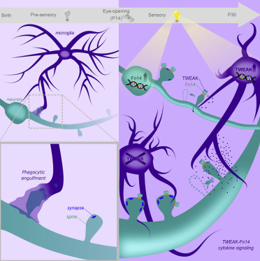
Graphical Abstract


Microglia affect the density of neuronal connections. CREDIT Lucas Cheadle


Highlights
• Experience induces Fn14 expression in neurons and TWEAK expression in microglia

• Fn14 increases the number of spine-associated synapses when not bound by TWEAK

• Microglial TWEAK signals through neuronal Fn14 to locally decrease synapse numbers

• Microglia-driven synapse loss occurs through a non-phagocytic mechanism

Summary
Sensory experience remodels neural circuits in the early postnatal brain through mechanisms that remain to be elucidated. Applying a new method of ultrastructural analysis to the retinogeniculate circuit, we find that visual experience alters the number and structure of synapses between the retina and the thalamus. These changes require vision-dependent transcription of the receptor Fn14 in thalamic relay neurons and the induction of its ligand TWEAK in microglia. Fn14 functions to increase the number of bulbous spine-associated synapses at retinogeniculate connections, likely contributing to the strengthening of the circuit that occurs in response to visual experience. However, at retinogeniculate connections near TWEAK-expressing microglia, TWEAK signals via Fn14 to restrict the number of bulbous spines on relay neurons, leading to the elimination of a subset of connections. Thus, TWEAK and Fn14 represent an intercellular signaling axis through which microglia shape retinogeniculate connectivity in response to sensory experience.

Authors
Lucas Cheadle, Samuel A. Rivera, Jasper S. Phelps, Katelin A. Ennis, Beth Stevens, Linda C. Burkly, Wei-Chung Allen Lee and Michael E. Greenberg.

Acknowledgements
The authors declare no competing financial interests. This is an open-access article distributed under the terms of the Creative Commons Attribution 4.0 International license, which permits unrestricted use, distribution and reproduction in any medium provided that the original work is properly attributed.

Brann along with Dr. Ratna Vadlamudi, Molecular Biologist, Professor of Obstetrics and Gynecology, University of Texas Health, San Antonio, are Co-corresponding Authors of the study and Co-principal Investigators on the National Institute of Neurological Disorders and Stroke grant funding the research (Grant R01NS088058).

Return to top of page.

Sep 17 2020   Fetal Timeline   Maternal Timeline   News
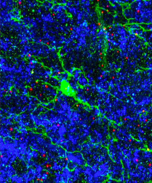


Microglia affect the density of neuronal connections. Microglial cells (stained in green), the immune cells of the mouse brain, send branches into areas with many neuronal connections (stained blue). Microglia make an immune signal, or cytokine, called TWEAK at the ends of their branches (stained in red). Microglia that contain large amounts of TWEAK (are deeper red) contact fewer neuronal connections (seen as black areas without blue connections). CREDIT Lucas Cheadle


Phospholid by Wikipedia