|
|
Developmental Biology - Placenta
Placenta Forms Days Before Embryo Begins
Placenta cells are initiated first, before cells of the fertilised egg divide and specialise...
The first stages of placental development take place days before the embryo starts to form in human pregnancies. This finding highlights the importance of a healthy placental development in pregnancy and could lead to future improvements in IVF fertility treatments and a better understanding of placental-related diseases in pregnancy.
In a study published in the journal Nature, researchers looked at the biological pathways active in human embryos during their first few days of development to understand how cells acquire different fates and functions within the early embryo.
Researchers observed that shortly after fertilisation and as cells start to divide, some cells stick together. This triggers a cascade of molecular events which initiate placental development. A subset of those cells change shape, or 'polarize', driving cells to become placental progenitor cells - precursor cells to a specialised placenta cell, which can be distinguished by differences in genes and proteins from other cells in the embryo.
"This study highlights the critical importance of the placenta for healthy human development. If the molecular mechanism we discovered for this first cell decision in humans is not appropriately established, this will have significant negative consequences for the development of the embryo and its ability to successfully implant in the womb."
Kathy K. Niakan PhD,
Centre for Trophoblast Research, Department of Physiology, Development and Neuroscience, University of Cambridge, Cambridge, UK;
Human Embryo and Stem Cell Laboratory, The Francis Crick Institute, London, UK.
The team also examined the same developmental pathways in mouse and cow embryos. They found that while the mechanisms of later stages of development differ between species, the placental progenitor is still the first cell to differentiate.
"We've shown that one of the earliest cell decisions during development is widespread in mammals, and this will help form the basis of future developmental research. Now we must continue to interrogate these pathways and identify biomarkers that facilitate healthy placental development in people, cows and other domestic animals.
Understanding the process of early human development in the womb could provide us with insights that may lead to improvements in IVF success rates in the future. It could also allow us to understand early placental dysfunctions that can pose a risk to human health later in pregnancy."
Claudia Gerri, lead author of the study and postdoctoral training fellow in the Human Embryo and Stem Cell Laboratory at the Francis Crick Institute.
During IVF, one of the most significant predictors of an embryo implanting in the womb is the appearance of placental progenitor cells under the microscope. If researchers could identify better markers of placental health or find ways to improve it, this could make a difference for people struggling to conceive.
This research was led by scientists at the Francis Crick Institute, in collaboration with colleagues at the Royal Veterinary College, Vrije Universiteit Brussel, Universite de Nantes and Bourn Hall Clinic. Kathy Niakan is incoming Director of the University of Cambridge's Centre for Trophoblast Research, and Chair of the Cambridge Strategic Research Initiative in Reproduction.
Human embryos examined during this study were in the morula stage of early-development, consisting of around 16-32 cells before progressing into the blastocyst stage (around 200 cells). They had been donated to research with consent by people undergoing in vitro fertilisation (IVF) and were not needed during the course of their treatment.
Abstract
Current understandings of cell specification in early mammalian pre-implantation development are based mainly on mouse studies. The first lineage differentiation event occurs at the morula stage, with outer cells initiating a trophectoderm (TE) placental progenitor program. The inner cell mass arises from inner cells during subsequent developmental stages and comprises precursor cells of the embryo proper and yolk sac1. Recent gene-expression analyses suggest that the mechanisms that regulate early lineage specification in the mouse may differ in other mammals, including human2,3,4,5 and cow6. Here we show the evolutionary conservation of a molecular cascade that initiates TE segregation in human, cow and mouse embryos. At the morula stage, outer cells acquire an apical?basal cell polarity, with expression of atypical protein kinase C (aPKC) at the contact-free domain, nuclear expression of Hippo signalling pathway effectors and restricted expression of TE-associated factors such as GATA3, which suggests initiation of a TE program. Furthermore, we demonstrate that inhibition of aPKC by small-molecule pharmacological modulation or Trim-Away protein depletion impairs TE initiation at the morula stage. Our comparative embryology analysis provides insights into early lineage specification and suggests that a similar mechanism initiates a TE program in human, cow and mouse embryos.
Authors
Claudia Gerri, Afshan McCarthy, Gregorio Alanis-Lobato, Andrej Demtschenko, Alexandre Bruneau, Sophie Loubersac, Norah M. E. Fogarty, Daniel Hampshire, Kay Elder, Phil Snell, Leila Christie, Laurent David, Hilde Van de Velde, Ali A. Fouladi-Nashta and Kathy K. Niakan.
Acknowledgements
The work was funded by the UK Medical Research Council, Wellcome and Cancer Research UK. It was approved by the UK Human Fertilisation and Embryology Authority (HFEA) and the Health Research Authority's Research Ethics Committee.
The authors thank the donors whose contributions have enabled this research; P. Patel, A. Srikantharajah, M. Summers, A. Handyside, K. Ahuja, S. Lavery, A. Rattos and M. Jansa Perez for the coordination and donation of embryos to our research project; members of the laboratories of K.K.N., J. M. A. Turner and R. Lovell-Badge, as well as M. Marass, T. Rayon Alonso, B. Thompson and N. Goehring for discussion, advice and feedback on the manuscript; the laboratories of B. Thompson, N. Goehring and J. Briscoe for sharing reagents and advice; the Advanced Light Microscopy and Biological Research Facilities (Francis Crick Institute); and A. Brodie and K. Bacon for assisting with cow embryo culture. The AMOT antibody (Amot-C no. 10061-1) used in this paper for mouse embryos was provided by the laboratory of H. Sasaki. CRT0276121 was provided by Cancer Research Technology. Work in the laboratory of L.D. was funded by a donation from MSD to ?Fondation de l?Universit? de Nantes?. Work in the laboratory of H.V.d.V. was funded by the Fonds Wetenschappelijk Onderzoek Flanders (FWOAL722) and the Wetenschappelijk Fonds Willy Gepts (WFWG, UZ Brussel, G142). Work in the laboratory of A.A.F.-N. was supported by Comparative Biomedical Sciences Departmental fund from the Royal Veterinary College. Work in the laboratory of K.K.N. was supported by the Francis Crick Institute, which receives its core funding from Cancer Research UK (FC001120), the UK Medical Research Council (FC001120) and the Wellcome Trust (FC001120), and by the Rosa Beddington Fund.
This study was approved by the UK Human Fertilisation and Embryology Authority (HFEA): research licence number R0162, and the Health Research Authority's Research Ethics Committee (Cambridge Central reference number 19/EE/0297).
Return to top of page.
|
|
Oct 1 2020 Fetal Timeline Maternal Timeline News
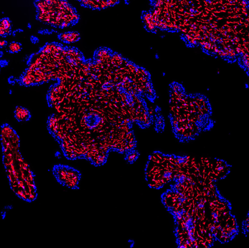
 Shortly after fertilisation as cells start to divide, some cells start to stick together. This triggers a cascade of molecular events that initiate placental development. CREDIT Rockefeller University/ Medical Express.
| |


