|
|
Developmental Biology - How Ovaries Maintain Fertility
An "Operating Manual" for Human Egg Fertility
Immature cells in ovaries go through a few steps to mature and maintain eggs...
Recently published work from the Carnegie Institution for Science has revealed, in unprecedented detail, the genetic steps immature egg cells go through as they mature, begin functioning and continue to maintain fertility.
The process of how immature egg cells are assisted by specific ovarian helper cells in the developing fetus, is well understood. But now, Allan Spradling and Howard Niu of The Carnegie Institute Department of Embryology, have mapped gene activity in thousands of these cells to learn how the stage is set for fertility later in women's lives.
Before birth, a female's "germ cells" assemble into a finite number of cell clusters called follicles located inside the female ovaries. Follicles are immature egg cells that act as "helper" cells to guide an egg through its maturation process. Mature egg cells will then burst away from these follicle cells in a process known as ovulation.
"Follicle cells are slowly used up during a female's reproductive lifespan. Menopause begins when they run out. Understanding what it takes for follicles to form and develop successfully, helps us learn how damaged genes or adverse environmental factors - like poor diet - can interfere with fertility.
Documenting follicle genetics creates an operating manual, where problems in egg development that lead to birth defects - from mutations or bad nutrition - can be better understood and reduced."
Allan C. Spradling PhD, Howard Hughes Medical Institute Research Laboratories; Department of Embryology, Carnegie Institution for Science, Baltimore, Maryland, USA.
Spradling and Niu sequenced 52,500 mouse ovarian cells at each of seven stages of follicle development, in order to determine the relative expression of thousands of follicular genes and to characterize the role each of these genes plays.
Their study illuminated how mammalian ovaries produce two distinct types of follicles and led them to be able to identify many differences in gene activity between them.
First - wave 1 follicles are present in the ovary even before puberty. In mice, these follicle cells generate the first fertile eggs. But, their function in humans is poorly understood. However, they may produce useful hormones.
The second type, called "wave 2 follicles", are stored in a resting state. But, small groups are activated to mature during a female's hormonal cycle, which ends in ovulation. These findings help clarify each cell type's role during this process.
Spradling and Niu's work and all its underlying data were published by PNAS, Proceedings of the National Academy of Sciences.
"We hope our work will serve as a genetic resource for all researchers who study reproduction and fertility."
Allan Spradling PhD and Wanbao Niu PhD, Howard Hughes Medical Institute Research Laboratories, Carnegie Institution for Science;
Department of Embryology, Carnegie Institution for Science, Baltimore, Maryland, USA.
Significance
This paper improves knowledge of the somatic and germ cells of the developing mouse ovary that assemble into ovarian follicles, by determining cellular gene expression, and tracing lineage relationships. The study covers the last week of fetal development through the first five days of postnatal development. During this time, many critically important processes take place, including sex determination, follicle assembly, and the initial events of meiosis. We report expression differences between pregranulosa cells of wave 1 follicles that function at puberty and wave 2 follicles that sustain fertility. These studies illuminate ovarian somatic cells and provide a resource to study the development, physiology, and evolutionary conservation of mammalian ovarian follicle formation.
Abstract
We sequenced more than 52,500 single cells from embryonic day 11.5 (E11.5) postembryonic day 5 (P5) gonads and performed lineage tracing to analyze primordial follicles and wave 1 medullar follicles during mouse fetal and perinatal oogenesis. Germ cells clustered into six meiotic substages, as well as dying/nurse cells. Wnt-expressing bipotential precursors already present at E11.5 are followed at each developmental stage by two groups of ovarian pregranulosa (PG) cells. One PG group, bipotential pregranulosa (BPG) cells, derives directly from bipotential precursors, expresses Foxl2 early, and associates with cysts throughout the ovary by E12.5. A second PG group, epithelial pregranulosa (EPG) cells, arises in the ovarian surface epithelium, ingresses cortically by E12.5 or earlier, expresses Lgr5, but delays robust Foxl2 expression until after birth. By E19.5, EPG cells predominate in the cortex and differentiate into granulosa cells of quiescent primordial follicles. In contrast, medullar BPG cells differentiate along a distinct pathway to become wave 1 granulosa cells. Reflecting their separate somatic cellular lineages, second wave follicles were ablated by diptheria toxin treatment of Lgr5-DTR-EGFP mice at E16.5 while first wave follicles developed normally and supported fertility. These studies provide insights into ovarian somatic cells and a resource to study the development, physiology, and evolutionary conservation of mammalian ovarian follicles.
Acknowledgments
The authors thank the Johns Hopkins University School of Medicine Biotechnology center for assistance with some of the scRNAseq experiments. We especially thank Allison Pinder and Fred Tan of the Carnegie Embryology Biotechnology Center for assistance in carrying out scRNAseq and in data analysis. We thank Mike Sepanski for carrying out electron microscopy. We thank Dr. Frederic J. de Sauvage (Genentech, Inc.) for kindly providing us with Lgr5-DTR-EGFP mice.
Return to top of page.
| |
|
Nov 18 2020 Fetal Timeline Maternal Timeline News
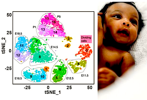
 Illustration of genes being expressed in waves 1 and 2 (ORANGE) of follicle production. Each dot on this diagram mathematically summarizes those genes expressed by an individual ovarian helper cell as it surrounds a developing egg cell. Developing egg cells are indicated by a single color and exist in the ovary only at specific times during development - E14.5, E16.5, etc., indicating their time zones. Each time zone houses two types of follicle cells, future wave 1 follicles (ODD #s) or wave 2 follicles (EVEN #s). CREDIT Allan Spradling & Wanbao Niu. Baby image - Shutterstock. Composite by Navid Marvi.
|


