|
|
Welcome to The Visible Embryo, a comprehensive educational resource on human development from conception to birth.
The Visible Embryo provides visual references for changes in fetal development throughout pregnancy and can be navigated via fetal development or maternal changes.
The National Institutes of Child Health and Human Development awarded Phase I and Phase II Small Business Innovative Research Grants to develop The Visible Embryo. Initally designed to evaluate the internet as a teaching tool for first year medical students, The Visible Embryo is linked to over 600 educational institutions and is viewed by more than one million visitors each month.
Today, The Visible Embryo is linked to over 600 educational institutions and is viewed by more than 1 million visitors each month. The field of early embryology has grown to include the identification of the stem cell as not only critical to organogenesis in the embryo, but equally critical to organ function and repair in the adult human. The identification and understanding of genetic malfunction, inflammatory responses, and the progression in chronic disease, begins with a grounding in primary cellular and systemic functions manifested in the study of the early embryo.

The World Health Organization (WHO) has created a new Web site to help researchers, doctors and patients obtain reliable information on high-quality clinical trials. Now you can go to one website and search all registers to identify clinical trial research underway around the world!

|
|
| Disclaimer: The Visible Embryo web site is provided for your general information only. The information contained on this site should not be treated as a substitute for medical, legal or other professional advice. Neither is The Visible Embryo responsible or liable for the contents of any websites of third parties which are listed on this site. |
|
|

Content protected under a Creative Commons License. Commons License. |
|
| No dirivative works may be made or used for commercial purposes. |
|
|
| |
|
|
 
CLICK ON weeks 0 - 40 and follow along every 2 weeks of fetal development
|
|
|
|
What happens if DNA cannot fit into the nucleus?
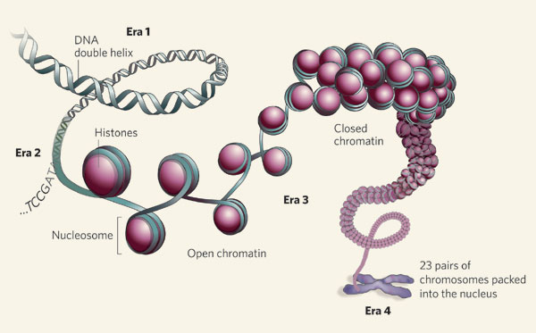
Packaging approximately 1.8 meters of DNA into something as small as a cell nucleus is no easy feat. But what about unpacking it again in order to access it's genes? All this activity requires organization. DNA is dynamic information that requires storage, access and constant maintenance.
In a nutshell, this is achieved through DNA being condensed and packaged in a complex of proteins called histones - or the chromatin structure. Histones are constantly being modified, the DNA is wound tightly to fit into the nucleus — and then must be "unwound" to be read and expressed as proteins. So, histones need constant replacement to maintain their correct structure in all intra-cellular DNA processes.
To understand more about histone replacement, researchers at the Babraham Institute and MRC Clinical Sciences Centre studied developing mouse eggs or oocytes. Developing eggs provide a system where the mechanics of how DNA is packaged into cells can be explored without being interferred with by DNA replication — as egg cells do not divide until fertilized by sperm.
Before the mature egg is ready to be fertilized, it's genome is still actively turning on and off DNA and genes. However, the work published in the latest issue of Molecular Cell, relied on the Institute's prior expertise in single cell analysis having mapped the epigenetic landscape of eggs.
These researchers were able to delete a single histone protein — one of the chaperone proteins responsible for replacing histones in chromatin — and analysed its effect on egg development, DNA integrity and methylation — that process by which methyl groups attach to DNA in order to modify it's function.
"Oocytes lacking the Hira histone, showed severe developmental defects which often led to cell death.
"The whole system was disrupted, eggs accumulated DNA damage and the altered chromatin [combination of DNA and histones] meant genes could not be either efficiently silenced or activated.
"But this uncovered an intricate relationship between different epigenetic systems operating in the oocyte. Failure to ensure normal histone levels severely compromised deposition of methyl groups onto DNA."
Gavin Kelsey PhD, group leader the Epigenetics Program, Babraham Institute, Cambridge, UK; and Centre for Trophoblast Research, University of Cambridge, Cambridge, UK
The research identified the relevance of histone turnover in maintaining gene precision, which adds to our appreciation of protecting the integrity of the genome as it is remodelled and reshaped.
In the context of the developing egg, this information reveals how important maintaining chromatin is to the integrity of egg formation.
Abstract Highlights
•Histone H3/H4 replacement is continuous and mediated by Hira during mouse oogenesis
•Loss of Hira results in chromatin abnormalities and extensive oocyte loss
•Hira depletion reduces histone load, which prevents normal transcriptional regulation
•Hira-mediated histone replacement is required for normal 5mC deposition in oocytes
Summary
The integrity of chromatin, which provides a dynamic template for all DNA-related processes in eukaryotes, is maintained through replication-dependent and -independent assembly pathways. To address the role of histone deposition in the absence of DNA replication, we deleted the H3.3 chaperone Hira in developing mouse oocytes. We show that chromatin of non-replicative developing oocytes is dynamic and that lack of continuous H3.3/H4 deposition alters chromatin structure, resulting in increased DNase I sensitivity, the accumulation of DNA damage, and a severe fertility phenotype. On the molecular level, abnormal chromatin structure leads to a dramatic decrease in the dynamic range of gene expression, the appearance of spurious transcripts, and inefficient de novo DNA methylation. Our study thus unequivocally shows the importance of continuous histone replacement and chromatin homeostasis for transcriptional regulation and normal developmental progression in a non-replicative system in vivo.
This is an open access article under the CC BY-NC-ND license (http://creativecommons.org/licenses/by-nc-nd/4.0/).
This work was supported by funding from the Biotechnology and Biological Research Council and Medical Research Council (MRC) to Gavin Kelsey at the Babraham Institute and the MRC and FP7 EpiGeneSys network to Petra Hajkova at the MRC Clinical Sciences Centre.
Return to top of page
|
|
|
Nov 10, 2015 Fetal Timeline Maternal Timeline News News Archive

Nucleosome structure incoporates DNA, Histone Core and Histone
Image Credit:
Nature Genomic architecture, and eras of investigation.
|
|
| |
|


