Welcome to The Visible Embryo
  o
 
The Visible Embryo Home
   
Google  
Home--- -History-----Bibliography-----Pregnancy Timeline-----Prescription Drugs in Pregnancy---- Pregnancy Calculator----Female Reproductive System----News----Contact
 
WHO International Clinical Trials Registry Platform


The World Health Organization (WHO) has a Web site to help researchers, doctors and patients obtain information on clinical trials. Now you can search all such registers to identify clinical trial research around the world!


Home

History

Bibliography

Pregnancy Timeline

Prescription Drug Effects on Pregnancy

Pregnancy Calculator

Female Reproductive System

News

Disclaimer: The Visible Embryo web site is provided for your general information only. The information contained on this site should not be treated as a substitute for medical, legal or other professional advice. Neither is The Visible Embryo responsible or liable for the contents of any websites of third parties which are listed on this site.


Content protected under a Creative Commons License.
No dirivative works may be made or used for commercial purposes.


Pregnancy Timeline by SemestersDevelopmental TimelineFertilizationFirst TrimesterSecond TrimesterThird TrimesterFirst Thin Layer of Skin AppearsEnd of Embryonic PeriodEnd of Embryonic PeriodFemale Reproductive SystemBeginning Cerebral HemispheresA Four Chambered HeartFirst Detectable Brain WavesThe Appearance of SomitesBasic Brain Structure in PlaceHeartbeat can be detectedHeartbeat can be detectedFinger and toe prints appearFinger and toe prints appearFetal sexual organs visibleBrown fat surrounds lymphatic systemBone marrow starts making blood cellsBone marrow starts making blood cellsInner Ear Bones HardenSensory brain waves begin to activateSensory brain waves begin to activateFetal liver is producing blood cellsBrain convolutions beginBrain convolutions beginImmune system beginningWhite fat begins to be madeHead may position into pelvisWhite fat begins to be madePeriod of rapid brain growthFull TermHead may position into pelvisImmune system beginningLungs begin to produce surfactant
CLICK ON weeks 0 - 40 and follow along every 2 weeks of fetal development


Neurons must wriggle to reach their final destination

As our brain develops, microtubules give nerve cells a boost along their way. With attachment help from motor proteins, microtubules send neurons from their birthplace to make a trip towards their final location. Once there, they pop out axons and dendrites to receive and send sensory signals.


Humans' most basic motor functions as well as cognitive thoughts, depend on this journey of neurons and the connections they make. A new study from Drexel University shows that the gliding movement of a small group of cellular structures called microtubules, play a key role in keeping neurons on their proper trajectory.

This discovery could ultimately help researchers better understand how neurons gone astray contribute to neurodevelopmental disorders, according to Peter Baas PhD, a professor in the Department of Neurobiology and Anatomy at Drexel University, College of Medicine in Philadelphia, Pennsylvania, USA and the study's principal investigator.


"This study is important for understanding how a healthy brain is organized. If neurons do not know when to start migrating, or where to go, or if the axons don't grow long enough, that can give way to disorders such as autism."

Peter Baas PhD, Department of Neurobiology and Anatomy, Drexel University College of Medicine, Philadelphia, PA, USA


The study, published this month in the Journal of Cell Biology, focuses on microtubules and the molecular motor proteins that generate force on these structures.

Until recently, the primary accepted idea was that microtubules' main functions were to grow longer and shorter — as "passive players" in the wiring of our nervous system. Researchers also assumed that all "functionally relevant" microtubules were attached to the centrosome, lying in the center of a cell's cytoplasm, near the nucleus. Called the organization center of a cell by Baas.

When a cell divides, each daughter cell receives one copy of the centrosome along with a pair of centrioles. We are used to seeing the centrioles depicted as lines pulling apart chromosomes during cell division.


Baas has spent his career studying the ways motor proteins push and pull on microtubules, enabling the axon to move in response to cues from the embryo.


Baas and his research team used electron tomography — an extension of traditional electron microscopy — to beam electrons through the center of a target at incremental degrees as the target is rotated. The data collected is reassembled into a three-dimensional image. Researchers wanted to see whether any microtubules detach from the centrosome, and if so, how detachment might affect how neurons travel.


Interplay between the centrosome cycle and the cell cycle
Journal of Cell Biology


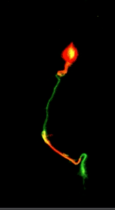
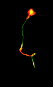
They found that a small group of microtubules are not attached to the centrosome, and that motor proteins can actually slide these unattached microtubules around within the neuron as it migrates.

Next, they wanted to know how those slithering, unattached microtubules mattered to how neurons move? So, they immobilized the motor protein ninein. Now, they saw that although neurons still slid around, they frequently changed direction, instead of moving in a simple, forward motion toward their intended goals.


"When we used the drug to inhibit sliding, we saw that the neuron can't migrate in a nice straight, smooth trajectory. That's how we found out that the little bit of sliding which normally occurs is really important for maneuverability."

Peter Baas PhD


Going a step further, researchers detached more microtubules from the centrosome by knocking out an anchoring protein. This caused many of the neurons to slow down or even come to a complete halt. Yet the neurons' axons continued to grow to long lengths.

By manipulating levels of protein, researchers now know that even the smallest alterations can greatly change the morphology and migratory behavior of a neuron, which can translate into developmental problems.


"If any of these mechanisms — ninein or any other motor protein — are disrupted, there can be problems."

Peter Baas PhD


Abstract
Contemporary models for neuronal migration are grounded in the view that virtually all functionally relevant microtubules (MTs) in migrating neurons are attached to the centrosome, which occupies a position between the nucleus and a short leading process. It is assumed that MTs do not undergo independent movements but rather transduce forces that enable movements of the centrosome and nucleus. The present results demonstrate that although this is mostly true, a small fraction of the MTs are centrosome-unattached, and this permits limited sliding of MTs. When this sliding is pharmacologically inhibited, the leading process becomes shorter, migration of the neuron deviates from its normal path, and the MTs within the leading process become buckled. Partial depletion of ninein, a protein that attaches MTs to the centrosome, leads to greater numbers of centrosome-unattached MTs as well as greater sliding of MTs. Concomitantly, the soma becomes less mobile and the leading process acquires an elongated morphology akin to an axon.

Return to top of page

May 23, 2016   Fetal Timeline   Maternal Timeline   News   News Archive   


By manipulating levels of the ninein protein, researchers watched as a neuron changed motion.
Normally creating a "wriggle" that appears to spur forward movement of a neuron, deleting the motor
protein ninein, caused neurons to lose forward momentum towards their intended goal.
Image Credit: Peter Baas, Drexel University Eurekalert


Phospholid by Wikipedia