|
|
Anatomy of a decision
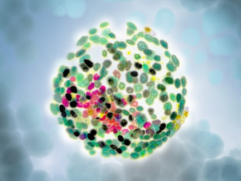
In the first genome-scale experiment of its kind, researchers have seen how a mouse embryo first transforms from a ball of unfocussed cells (a gastrula) into a structured embryo.
Published in Nature, the single-cell genomics study was led by the European Bioinformatics Institute (EMBL-EBI) and the Wellcome Trust-MRC Cambridge Stem Cell Institute.
Gastrulation is the point when an animal's whole body plan is set, just before individual organs start to develop. Understanding this point in very early development is vital to understanding how animals develop and how things go wrong. One of the biggest challenges in studying gastrulation is the very small number of cells that make up the embryo at this stage.
"If we want to better understand the natural world around us, one of the fundamental questions is, how do animals develop? How do you turn from an egg into an animal, with all sorts of tissues? Many of the things that go wrong, like birth defects, are caused by problems in early development. We need to have an atlas of normal development for comparison when things go wrong."
Bertie Gottgens PhD, Wellcome Trust - Medical Research Council Cambridge Stem Cell Institute, University of Cambridge, UK, and Research Group Leader.
Today, thanks to advances in single-cell sequencing, the team was able to analyse over 1000 individual cells of gastrulating mouse embryos. The result is an atlas of gene expression during very early, healthy mammalian development.
"Single-cell technologies are a major change over what we've used before - we can now make direct observations and see what's going on during the earliest stages of development.
"Once we have that information, we can take cells from embryos in which genetic factors are not working properly at a specific developmental stage, and map them to the healthy atlas to better understand what might be happening."
John Marioni, Research Group Leader at EMBL-EBI, the Wellcome Trust Sanger Institute, and the University of Cambridge.
To illustrate the usefulness of the atlas, the team studied what happened when they removed from blood, a genetic factor essential to formation of blood cells.
"It wasn't what we expected at all. We found cells — which in healthy embryos would commit to becoming blood cells — actually become confused in the embryos lacking that key gene. Effectively, getting stuck," explains Marioni. "What is so exciting is that we demonstrate how we can look at a very small number of cells at the precise moment a decision is being made. It gives us a completely different perspective on development."
Adds Gottgens: "This is just the beginning of how single cell genomics will transform our understanding of early development."
Abstract
In mammals, specification of the three major germ layers occurs during gastrulation, when cells ingressing through the primitive streak differentiate into the precursor cells of major organ systems. However, the molecular mechanisms underlying this process remain unclear, as numbers of gastrulating cells are very limited. In the mouse embryo at embryonic day 6.5, cells located at the junction between the extra-embryonic region and the epiblast on the posterior side of the embryo undergo an epithelial-to-mesenchymal transition and ingress through the primitive streak.
Subsequently, cells migrate, either surrounding the prospective ectoderm contributing to the embryo proper, or into the extra-embryonic region to form the yolk sac, umbilical cord and placenta. Fate mapping has shown that mature tissues such as blood and heart originate from specific regions of the pre-gastrula epiblast, but the plasticity of cells within the embryo and the function of key cell-type-specific transcription factors remain unclear.
Here we analyse 1,205 cells from the epiblast and nascent Flk1+ mesoderm of gastrulating mouse embryos using single-cell RNA sequencing, representing the first transcriptome-wide in vivo view of early mesoderm formation during mammalian gastrulation. Additionally, using knockout mice, we study the function of Tal1, a key haematopoietic transcription factor, and demonstrate, contrary to previous studies performed using retrospective assays, that Tal1 knockout does not immediately bias precursor cells towards a cardiac fate.
Return to top of page
|
|
|
Jul 19, 2016 Fetal Timeline Maternal Timeline News News Archive
The Wellcome Trust–Medical Research Council Cambridge Stem Cell Institute, UK -
has identified factors sparking the formation of pluripotent cells.
Image Credit: Maulik Patel, Vanderbilt University

(A) Overview of the developmental sequence analyzed.
Image Credit: MRC Cambridge Stem Cell Institute
|


