Welcome to The Visible Embryo
  o
 
The Visible Embryo Home
   
Google  
Home--- -History-----Bibliography-----Pregnancy Timeline-----Prescription Drugs in Pregnancy---- Pregnancy Calculator----Female Reproductive System----News----Contact
 
WHO International Clinical Trials Registry Platform


The World Health Organization (WHO) has a Web site to help researchers, doctors and patients obtain information on clinical trials. Now you can search all such registers to identify clinical trial research around the world!


Home

History

Bibliography

Pregnancy Timeline

Prescription Drug Effects on Pregnancy

Pregnancy Calculator

Female Reproductive System

News

Disclaimer: The Visible Embryo web site is provided for your general information only. The information contained on this site should not be treated as a substitute for medical, legal or other professional advice. Neither is The Visible Embryo responsible or liable for the contents of any websites of third parties which are listed on this site.


Content protected under a Creative Commons License.
No dirivative works may be made or used for commercial purposes.


Pregnancy Timeline by SemestersDevelopmental TimelineFertilizationFirst TrimesterSecond TrimesterThird TrimesterFirst Thin Layer of Skin AppearsEnd of Embryonic PeriodEnd of Embryonic PeriodFemale Reproductive SystemBeginning Cerebral HemispheresA Four Chambered HeartFirst Detectable Brain WavesThe Appearance of SomitesBasic Brain Structure in PlaceHeartbeat can be detectedHeartbeat can be detectedFinger and toe prints appearFinger and toe prints appearFetal sexual organs visibleBrown fat surrounds lymphatic systemBone marrow starts making blood cellsBone marrow starts making blood cellsInner Ear Bones HardenSensory brain waves begin to activateSensory brain waves begin to activateFetal liver is producing blood cellsBrain convolutions beginBrain convolutions beginImmune system beginningWhite fat begins to be madeHead may position into pelvisWhite fat begins to be madePeriod of rapid brain growthFull TermHead may position into pelvisImmune system beginningLungs begin to produce surfactant
CLICK ON weeks 0 - 40 and follow along every 2 weeks of fetal development


A molecule for making friends

The Urocortin-3 molecular helps mice and humans find new friends according to research from the Max Planck Institute of Psychiatry.


Meeting new people can be both stressful and rewarding — and may involve the same molecule. New research conducted in Alon Chen's Department, and led by Yair Shemesh and Oren Forkosh of the Max Planck Institute of Psychiatry, suggests Urocortin-3, a molecule which regulates stress — may also determine how willingly we leave our preferred social group to strike up new relationships.

Research results are published in the journal Nature Neuroscience.

Working with mice, researchers identified a stress mechanism that appears to act as a "social switch." It causes mice to either increase interactions with "friends" and "acquaintances" or reduce those interactions and seek to meet strangers instead. As a similar stress system operates in humans, their findings suggest a similar mechanism regulates social challenges in humans. Disruptions in this mechanism might be responsible for difficulties in people affected by social anxiety, as well as in those with autism, schizophrenia or other disorders.


"Most social contact involves a level of social stress or anxiety when we interact with people we know well, such as during a party with extended family. In fact, from the point of view of evolution, moderate levels of social apprehension are essential for safe and successful social engagement."

Yair Shemesh PhD, Department of Neurobiology, Weizmann Institute of Science, Rehovot, Israel; Department of Stress Neurobiology and Neurogenetics, Max Planck Institute of Psychiatry, Munich, Germany.

"In any social environment, an individual's interests often clash with the group's needs and expectations. So, the individual must maintain what's known as a socioemotional balance: between processing social signals and his or her emotional response to such pressures."

Alon Chen PhD, Department of Neurobiology, Weizmann Institute of Science, Rehovot, Israel; Department of Stress Neurobiology and Neurogenetics, Max Planck Institute Psychiatry, Munich, Germany.


Scientists used two behavioral setups to study how mice cope with the challenge of interacting with other mice. One, a "social maze," in which a mouse can choose to interact through a mesh with either familiar mice or strangers, or avoid interaction all together. The other was a special arena, in which mice were tracked with video cameras while observations were analyzed using a computer algorithm created for that purpose. This unique setup enabled researchers to quantify various types of interactions — approach, contact, attack or chase — among individual mice within the undisturbed group over several days.

The results revealed that a molecular mechanism involved in stress management in mice brains determines their behavior toward other mice. A small signaling molecule, Urocortin-3, attaches to a receptor on the surface of neurons, binding them to the molecule.


Both Urocortin-3 and it's receptor are part of the corticotropin-releasing factor, or CRF system. A system that plays a central role in coping with stress.

Both the CRF factor and receptor are prominently expressed in a brain region called the medial amygdala — a part of the brain in mice associated with social behavior.


Mice with high levels of Urocortin-3 in their brain actively seek out contacts with new mice behind the mesh, even ignoring their own group. But when the brain activity of Urocortin-3 and its receptor are blocked, the mice chose to socialize mainly within their group, avoiding contact with strangers.

Adds Oren Forkosh: "In nature, mice live in groups and the social challenges they face differ from their relationship with intruders. It therefore makes sense for a brain mechanism to produce different types of social coping in these two situations. In humans, this mechanism might be involved whenever we consider moving out of our parents' home, getting a divorce or changing jobs or apartments."

Abstract
Social encounters are associated with varying degrees of emotional arousal and stress. The mechanisms underlying adequate socioemotional balance are unknown. The medial amygdala (MeA) is a brain region associated with social behavior in mice. Corticotropin-releasing factor receptor type-2 (CRF-R2) and its specific ligand urocortin-3 (Ucn3), known components of the behavioral stress response system, are highly expressed in the MeA. Here we show that mice deficient in CRF-R2 or Ucn3 exhibit abnormally low preference for novel conspecifics. MeA-specific knockdown of Crfr2 (Crhr2) in adulthood recapitulated this phenotype. In contrast, pharmacological activation of MeA CRF-R2 or optogenetic activation of MeA Ucn3 neurons increased preference for novel mice. Furthermore, chemogenetic inhibition of MeA Ucn3 neurons elicited pro-social behavior in freely behaving groups of mice without affecting their hierarchal structure. These findings collectively suggest that the MeA Ucn3–CRF-R2 system modulates the ability of mice to cope with social challenges.

Original publication:
Yair Shemesh, Oren Forkosh, Mathias Mahn, Sergey Anpilov, Yehezkel Sztainberg, Sharon Manashirov, Tamar Shlapobersky, Evan Elliott, Laure Tabouy, Gili Ezra, Elaine S Adler, Yair J Ben-Efraim, Shosh Gil, Yael Kuperman, Sharon Haramati, Julien Dine, Matthias Eder, Jan M Deussing, Elad Schneidman, Ofer Yizhar & Alon Chen Ucn3 and CRF-R2 in the medial amygdala regulate complex social dynamics Nature Neuroscience; 18 July, 2016

Return to top of page

Jul 27, 2016   Fetal Timeline   Maternal Timeline   News   News Archive   
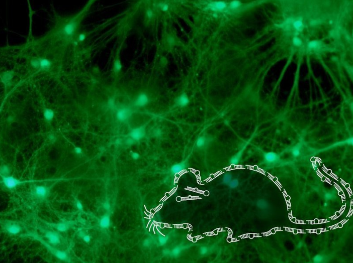


A specific molecule — Urocortin-3 — regulates stress-induced behavior in mice.
Image Credit:© Tali Wiesel /Weizmann Institute of Science.


Phospholid by Wikipedia