|
|
'Amplifier' helps make connections in the fetal brain
A special amplifier makes neural signals stronger in babies — then stops once neural connections are fully strengthened.
Fetal brains use a special amplifier to transmit signals, according to research by Matthew Colonnese PhD and Yasunobu Murata PhD of George Washington University (GW). Early neural connections are sparse, weak, and unreliable — so this unique amplification circuit boosts weak input to ensure accurate and powerful information transfer in the developing brain.
The study is published in the journal eLife.
"Our question is, what is the brain of the fetus doing? We know it's active, and we know it's generating spontaneous activity, but we also know that circuits are very weak.
"Brain activity in a pre-term infant is large - 10 times larger than that of an adult. At the same time, circuits have just ten percent of the connection of an adult.
"The question became how does the activity gets through? That's when we started looking for amplifiers and through our research, identified one."
Matthew Colonnese PhD, Professor, Pharmacology and Physiology, member Institute for Neuroscience, George Washington School of Medicine and Health Sciences, Washington, D.C., USA
Colonnese and Murata looked at key processing areas of the the cerebral cortex and thalamus in an animal model substituting the human fetal brain during development. The visual pathways of the animal model have similar activity patterns as humans.
In adults, activity in the eye is transmitted to the thalamus, then on to the cortex, without being amplified. Then feedback from the brain cortex back to the thalamus, modifies the signal strength — but mostly acts as a brake.
However, during early brain development feedback from the cortex multiplies retinal input to the thalamus without restraining it. This results in an excitatory or excited feedback loop which causes massive increases in activity. This amplifier explains how a fetal brain continues to stay active despite weak synapse connections.
While further research is needed for support and verification, the study may explain the differences in seizures between the young and old. In adults, this brake is needed to prevent runaway excitation which may cause epilepsy.
"The amplifier only lasts during the fetal period and then a brake comes on. The amplifier goes away after birth, but if it doesn't work or go away properly, does it cause disorders?
"This is likely one of many unique circuits the fetal brain is using. Research on the fetal brain is so new, and there is so much we don't yet know - it's like going to the moon.
"More research is needed to learn about overall fetal brain development and the significance of this amplifier."
Matthew Colonnese PhD
Abstract
Spontaneous retinal waves are critical for the development of receptive fields in visual thalamus (LGN) and cortex (VC). Despite a detailed understanding of the circuit specializations in retina that generate waves, whether central circuit specializations also exist to control their propagation through visual pathways of the brain is unknown. Here we identify a developmentally transient, corticothalamic amplification of retinal drive to thalamus as a mechanism for retinal wave transmission in the infant rat brain. During the period of retinal waves, corticothalamic connections excite LGN, rather than driving feedforward inhibition as observed in the adult. This creates an excitatory feedback loop that gates retinal wave transmission through the LGN. This cortical multiplication of retinal wave input ends just prior to eye-opening, as cortex begins to inhibit LGN. Our results show that the early retino-thalamo-cortical circuit uses developmentally specialized feedback amplification to ensure powerful, high-fidelity transmission of retinal activity despite immature connectivity.
"An excitatory cortical feedback loop gates retinal wave transmission in thalamus" was published in eLife and is available at http://dx.doi.org/10.7554/eLife.18816.
The work was supported by a grant from the National Eye Institute.
About the GW School of Medicine and Health Sciences: Founded in 1824, the GW School of Medicine and Health Sciences (SMHS) was the first medical school in the nation's capital and is the 11th oldest in the country. Working together in our nation's capital, with integrity and resolve, the GW SMHS is committed to improving the health and well-being of our local, national and global communities. smhs.gwu.edu
Return to top of page
|
|
|
Oct 12, 2016 Fetal Timeline Maternal Timeline News News Archive
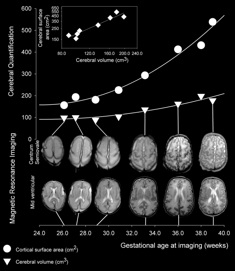
 Human cortical development between 26 and 39 week gestational age.
Image Credit: Wikipedia
|


