|
|||||||||||||||
|

CLICK ON weeks 0 - 40 and follow along every 2 weeks of fetal development
|
||||||||||||||||||||||||||||
Brainstem and visual cortex control our eyes "This study elegantly shows how the visual cortex continues to surprise and awe," said Houmam Araj PhD, a program director at the National Eye Institute (NEI). The study, published in Nature, was led by researchers at the University of California, San Diego (UCSD) and the University of California, San Francisco (UCSF) while funded by the National Eye Institute (NEI), part of the National Institutes of Health. Without our being aware of it, our eyes are in constant motion. As we rotate our heads, the world moves around us. Two ocular reflexes work to offset that head movement and stabilize any images being projected onto our retinas — the light-sensitive tissue at the back of our eyes. The two reflexes occur automatically as a result of signals from the brainstem, an evolutionarily older part of our brain. Both are precisely coordinated to each other, when one is impaired, the other compensates. This orchestration requires adaptive plasticity, says Massimo Scanziani PhD, the study's lead investigator who was a professor of neurobiology at UCSD at the time of the study, before moving to UCSF. Scanziani and his colleagues, first author Bao-hua Liu PhD - a post-doc at UCSD, and Andrew Huberman PhD - now associate professor at Stanford University, sought to understand the origins of adaptive plasticity by studying the eye movements in mice. In the mouse model, they disabled the vestibulo-ocular reflex which increases the optokinetic reflex. They measured reflex increase by keeping the mouse's head still while rotating visual stimuli in the form of black and white horizontal stripes around the head. Cameras recorded eye movements. More forceful eye movements indicated increased optokinetic reflex activity. To test the role of the visual cortex in the adaptive plasticity reflexes, they used a technique called optogenetics — which uses light to turn target cells on or off. Targeted cells were the inhibitory neurons in the visual cortex. Turning them "on" silences that region of the brain. It has long been observed that a collection of neural projections from the visual cortex extends into the brainstem which regulates innate motor behaviors. Lesioning these projections, a decrease occured in the optokinetic reflex. This suggests these neural projections are the anatomical structures with which the visual cortex adjusts plasticity in the optokinetic reflex.
Abstract The mammalian visual cortex massively innervates the brainstem, a phylogenetically older structure, via cortico-fugal axonal projections1. Many cortico-fugal projections target brainstem nuclei that mediate innate motor behaviours, but the function of these projections remains poorly understood1, 2, 3, 4. A prime example of such behaviours is the optokinetic reflex (OKR), an innate eye movement mediated by the brainstem accessory optic system3, 5, 6, that stabilizes images on the retina as the animal moves through the environment and is thus crucial for vision5. The OKR is plastic, allowing the amplitude of this reflex to be adaptively adjusted relative to other oculomotor reflexes and thereby ensuring image stability throughout life7, 8, 9, 10, 11. Although the plasticity of the OKR is thought to involve subcortical structures such as the cerebellum and vestibular nuclei10, 11, 12, 13, cortical lesions have suggested that the visual cortex might also be involved9, 14, 15. Here we show that projections from the mouse visual cortex to the accessory optic system promote the adaptive plasticity of the OKR. OKR potentiation, a compensatory plastic increase in the amplitude of the OKR in response to vestibular impairment11, 16, 17, 18, is diminished by silencing visual cortex. Furthermore, targeted ablation of a sparse population of cortico-fugal neurons that specifically project to the accessory optic system severely impairs OKR potentiation. Finally, OKR potentiation results from an enhanced drive exerted by the visual cortex onto the accessory optic system. Thus, cortico-fugal projections to the brainstem enable the visual cortex, an area that has been principally studied for its sensory processing function19, to plastically adapt the execution of innate motor behaviours. The study was supported in part by NEI grant R01 EY025668. Reference: Liu, Bao-hua et al. "Cortico-fugal output from visual cortex promotes plasticity of innate motor behavior." Published online in Nature, October 12, 2016. DOI:10.1038/nature19818 NEI leads the federal government's research on the visual system and eye diseases. NEI supports basic and clinical science programs that result in the development of sight-saving treatments. For more information, visit http://www.nei.nih.gov. About the National Institutes of Health (NIH): NIH, the nation's medical research agency, includes 27 Institutes and Centers and is a component of the U.S. Department of Health and Human Services. NIH is the primary federal agency conducting and supporting basic, clinical, and translational medical research, and is investigating the causes, treatments, and cures for both common and rare diseases. For more information about NIH and its programs, visit http://www.nih.gov. |
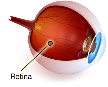
Oct 20, 2016 Fetal Timeline Maternal Timeline News News Archive  Human eye retina. Image Credit: Public Domain
|
||||||||||||||||||||||||||||

