Welcome to The Visible Embryo
  o
 
The Visible Embryo Home
   
Google  
Home--- -History-----Bibliography-----Pregnancy Timeline-----Prescription Drugs in Pregnancy---- Pregnancy Calculator----Female Reproductive System----News----Contact

   
WHO International Clinical Trials Registry Platform

The World Health Organization (WHO) has a Web site to help researchers, doctors and patients obtain information on clinical trials.

Now you can search all such registers to identify clinical trial research around the world!


Home

History

Bibliography

Pregnancy Timeline

Prescription Drug Effects on Pregnancy

Pregnancy Calculator

Female Reproductive System

News

Disclaimer: The Visible Embryo web site is provided for your general information only. The information contained on this site should not be treated as a substitute for medical, legal or other professional advice. Neither is The Visible Embryo responsible or liable for the contents of any websites of third parties which are listed on this site.


Content protected under a Creative Commons License.
No dirivative works may be made or used for commercial purposes.


Pregnancy Timeline by SemestersDevelopmental TimelineFertilizationFirst TrimesterSecond TrimesterThird TrimesterFirst Thin Layer of Skin AppearsEnd of Embryonic PeriodEnd of Embryonic PeriodFemale Reproductive SystemBeginning Cerebral HemispheresA Four Chambered HeartFirst Detectable Brain WavesThe Appearance of SomitesBasic Brain Structure in PlaceHeartbeat can be detectedHeartbeat can be detectedFinger and toe prints appearFinger and toe prints appearFetal sexual organs visibleBrown fat surrounds lymphatic systemBone marrow starts making blood cellsBone marrow starts making blood cellsInner Ear Bones HardenSensory brain waves begin to activateSensory brain waves begin to activateFetal liver is producing blood cellsBrain convolutions beginBrain convolutions beginImmune system beginningWhite fat begins to be madeHead may position into pelvisWhite fat begins to be madePeriod of rapid brain growthFull TermHead may position into pelvisImmune system beginningLungs begin to produce surfactant
CLICK ON weeks 0 - 40 and follow along every 2 weeks of fetal development


Earliest embryonic stem cells identified

Researchers have identified cell surface markers specific to the very earliest stem cells in a human embryo. These cells are believed to have the greatest potential in replacing damaged tissue. But until now, early stem cells have been difficult to distinguish from classic embryonic stem cells.


A new type of pluripotent stem cell genuinely corresponds to a more immature, pre-implantation stage, and has been identified by researchers at the Karolinska Institutet in Germany. These stem cells can now be cultivated in a laboratory and are of great scientific importance as they have the potential to build cell types difficult to create from classical stem cells. They may also be easier to cultivate and manipulate in laboratory experiments.

During the first week of fertilisation, an embryo grows from a single cell into a hollow cluster of a few hundred cells called a blastocyst. The blastocyst then attaches to the wall of the uterus in the stage called implantation.


For a limited period from fertilisation to a few days after implantation — an embryo contains pluripotent stem cells (or PSCs). PSC cells can develop into every cell type in the body, and are of considerable interest in regenerative and reparative medicine.


A few years ago, it was discovered there are two stages for human pluripotent stem cells, corresponding to pre-implanted and post-implanted embryonic cells. Although the classical stem cells used in regenerative medicine are isolated from a pre-implanted embryo, they reflect a mature stage more similar to a post-implantation embryo.

The work is published in the prestigious journal Cell Stem Cell.

Fredrik Lanner's research team at Karolinska Institutet and with their colleagues in Peter Rugg-Gunn's team at Cambridge's Babraham Institute in the United Kingdom, have now developed a tool for separating the two stem cell stages.

They have screened combinations of antibodies that bind to specific proteins on the surface of immature stem cells as well as antibodies that bind to specific proteins on surfaces of mature stem cells, which can be used in flow cytometry — a common laboratory technique used to sort cells.


"We've not had cell surface markers for different stem cell states before, making them hard to study. Now we have a simple tool for identifying and sorting cells, which benefits future stem cell research and basic research in early embryo development."

Fredrik Lanner PhD, Assistant Professor, Department of Clinical Science, Intervention and Technology, Karolinska Institute, Germany.


Mature embryonic stem cells cultivated in the laboratory can, under the right conditions, be 'backed up' in development to a more immature stem cell stage. Researchers tested their technique on cultivated stem cells from both mature and immature stages, and on donated human embryos left over from IVF treatments.


As expected, only the immature stem cell type was identified in pre-implanted embryos, which indicates that the antibodies are highly specific.


"It is at the point of implantation that the stem cells go through this change and 'mature', which is also a highly critical time for the embryo," says Dr Lanner. "These cells are therefore also of interest to infertility research."

Abstract
Human pluripotent stem cells (PSCs) exist in naive and primed states and provide important models to investigate the earliest stages of human development. Naive cells can be obtained through primed-to-naive resetting, but there are no reliable methods to prospectively isolate unmodified naive cells during this process. Here we report comprehensive profiling of cell surface proteins by flow cytometry in naive and primed human PSCs. Several naive-specific, but not primed-specific, proteins were also expressed by pluripotent cells in the human preimplantation embryo. The upregulation of naive-specific cell surface proteins during primed-to-naive resetting enabled the isolation and characterization of live naive cells and intermediate cell populations. This analysis revealed distinct transcriptional and X chromosome inactivation changes associated with the early and late stages of naive cell formation. Thus, identification of state-specific proteins provides a robust set of molecular markers to define the human PSC state and allows new insights into the molecular events leading to naive cell resetting.

Highlights
• Flow cytometry profiles cell surface proteins in naive and primed human PSCs
• The human PSC state can be defined using robust state-specific protein markers
• Identified cell surface proteins track the dynamics of naive-primed PSC conversions
•Analyses of early-stage naive cells reveal transcription events during conversion

The study was financed by several bodies, including the Swedish Research Council, the Ragnar Söderberg Foundation, the Swedish Foundation for Strategic Research, the Knut and Alice Wallenberg Foundation, the Centre for Innovative Medicine (CIMED) and the Ming Wai Lau Centre for Reparative Medicine.

Publication: 'Comprehensive Cell Surface Protein Profiling Identifies Specific Markers of Human Naive and Primed Pluripotent States', Amanda J. Collier, Sarita P. Panula, John Paul Schell, Peter Chovanec, Alvaro Plaza Reyes, Sophie Petropoulos, Anne E. Corcoran, Rachael Walker, Iyadh Douagi, Fredrik Lanner, Peter J. Rugg-Gunn. Cell Stem Cell, online 23 March 2017, doi: 10.1016/j.stem.2017.02.014.
Return to top of page

Apr 5, 2017   Fetal Timeline   Maternal Timeline   News   News Archive   
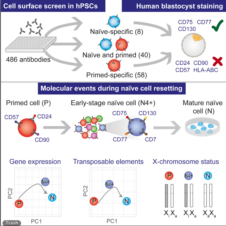


Screening for antigens that identify immature from mature stem cells.
Image Credit: Fredrik Lanner, Karolinska Institutet


Phospholid by Wikipedia