Welcome to The Visible Embryo

Home-- -History-- -Bibliography- -Pregnancy Timeline- --Prescription Drugs in Pregnancy- -- Pregnancy Calculator- --Female Reproductive System- -Contact
 

Welcome to The Visible Embryo, a comprehensive educational resource on human development from conception to birth.

The Visible Embryo provides visual references for changes in fetal development throughout pregnancy and can be navigated via fetal development or maternal changes.

The National Institutes of Child Health and Human Development awarded Phase I and Phase II Small Business Innovative Research Grants to develop The Visible Embryo. Initally designed to evaluate the internet as a teaching tool for first year medical students, The Visible Embryo is linked to over 600 educational institutions and is viewed by more than one million visitors each month.

Today, The Visible Embryo is linked to over 600 educational institutions and is viewed by more than 1 million visitors each month. The field of early embryology has grown to include the identification of the stem cell as not only critical to organogenesis in the embryo, but equally critical to organ function and repair in the adult human. The identification and understanding of genetic malfunction, inflammatory responses, and the progression in chronic disease, begins with a grounding in primary cellular and systemic functions manifested in the study of the early embryo.

WHO International Clinical Trials Registry Platform


The World Health Organization (WHO) has created a new Web site to help researchers, doctors and
patients obtain reliable information on high-quality clinical trials. Now you can go to one website and search all registers to identify clinical trial research underway around the world!


Home

History

Bibliography

Pregnancy Timeline

Prescription Drug Effects on Pregnancy

Pregnancy Calculator

Female Reproductive System

Contact The Visible Embryo

News Alerts Archive

Disclaimer: The Visible Embryo web site is provided for your general information only. The information contained on this site should not be treated as a substitute for medical, legal or other professional advice. Neither is The Visible Embryo responsible or liable for the contents of any websites of third parties which are listed on this site.
Content protected under a Creative Commons License.

No dirivative works may be made or used for commercial purposes.

Return To Top Of Page
Pregnancy Timeline by SemestersFetal liver is producing blood cellsHead may position into pelvisBrain convolutions beginFull TermWhite fat begins to be madeWhite fat begins to be madeHead may position into pelvisImmune system beginningImmune system beginningPeriod of rapid brain growthBrain convolutions beginLungs begin to produce surfactantSensory brain waves begin to activateSensory brain waves begin to activateInner Ear Bones HardenBone marrow starts making blood cellsBone marrow starts making blood cellsBrown fat surrounds lymphatic systemFetal sexual organs visibleFinger and toe prints appearFinger and toe prints appearHeartbeat can be detectedHeartbeat can be detectedBasic Brain Structure in PlaceThe Appearance of SomitesFirst Detectable Brain WavesA Four Chambered HeartBeginning Cerebral HemispheresFemale Reproductive SystemEnd of Embryonic PeriodEnd of Embryonic PeriodFirst Thin Layer of Skin AppearsThird TrimesterSecond TrimesterFirst TrimesterFertilizationDevelopmental Timeline
CLICK ON weeks 0 - 40 and follow along every 2 weeks of fetal development
Google Search artcles published since 2007


Home | Pregnancy Timeline | News Alerts |News Archive Aug 9, 2013


The structure of NatA, an n-terminal acetyltransferase.

Image credit: The Wistar Institute/Glen Liszczak


WHO Child Growth Charts


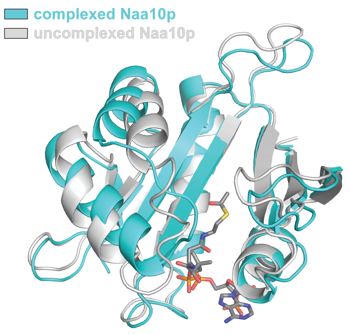
NatA complex may be a target for cancer treatment

NatA is an enzyme complex critical to cell growth. It's production is also elevated in many cancers, making it a target for tumor therapy.

A study in Nature Structural & Molecular Biology, led by researchers at The Wistar Institute, depicts the structure and the means of action of a protein complex called NatA. Their findings, they believe, will allow them to create an inhibitor—a potential drug—that could knock out NatA in order to curb the growth of cancer cells.


"NatA is essential for the growth of cells and their ability to divide, and we can see elevated production of this enzyme in many forms of cancer. Obviously, this is a particularly appealing drug target and we are currently leveraging our recent understanding of how the protein works to develop small molecules that will bind to and inactivate NatA."

Ronen Marmorstein, Ph.D., senior author, Hilary Koprowski, M.D. Professor, and leader of The Wistar Institute Cancer Center's Gene Expression and Regulation program.


NatA is a member of a family of N-terminal acetyltransferase (NAT) enzymes (or enzyme complexes) that modify proteins in order to control their behavior—for example by turning proteins on, telling proteins where to move, and tagging proteins or the cell for destruction.

According to Marmorstein, NatA works with amazing specificity for a particular sequence of amino acids—the individual building blocks of proteins—and by unraveling the roots of that specificity, the scientists have solved an alluring puzzle.

The Marmorstein laboratory has a long history studying acetylation enzymes, proteins that modify other molecules in the cell by adding an acetyl "tag."


In the cellular world, structure dictates function, and acetylation is a universal process for controlling protein behavior and gene expression in living organisms.

Marmorstein explaines: "Modifying protein structures is one way that our cells control how proteins function, and enzymes in the NAT family modify nearly 85 percent of human proteins, while NatA modifies 50 percent of these."


According to Marmorstein, NatA operates in a complex of two proteins, an enzyme subunit and it's auxiliary partner. When they worked out the structure of NatA—by bombarding a crystallized sample of the enzyme with powerful X-rays—they found how the auxiliary partner is crucial to turning the enzymatic subunit on.


Binding an auxiliary protein causes structural change to the enzyme subunit, configuring an active site on the protein—a region where chemical reaction can occur—turning it into a "switch" that activates the enzyme.


"When it binds to its auxiliary protein, the enzymatic subunit of NatA actually changes shape, reconfiguring the structure to allow it to properly grab onto its target's protein N-terminal sequence for acetylation," Marmorstein adds.

Importantly, others have found that NatA function is required for the proliferation of cancer cells. Marmorstein believes understanding the structure of NatA has allowed his team to better understand how to inactivate the protein in cancer cells.

The structure has yielded targets for small molecules that will act as inhibitors, essentially stopping the protein by gumming up its structure.

Abstract
N-terminal acetylation is ubiquitous among eukaryotic proteins and controls a myriad of biological processes. Of the N-terminal acetyltransferases (NATs) that facilitate this cotranslational modification, the heterodimeric NatA complex has the most diversity for substrate selection and modifies the majority of all N-terminally acetylated proteins. Here, we report the X-ray crystal structure of the 100-kDa holo-NatA complex from Schizosaccharomyces pombe, in the absence and presence of a bisubstrate peptide-CoA–conjugate inhibitor, as well as the structure of the uncomplexed Naa10p catalytic subunit. The NatA-Naa15p auxiliary subunit contains 13 tetratricopeptide motifs and adopts a ring-like topology that wraps around the NatA-Naa10p subunit, an interaction that alters the Naa10p active site for substrate-specific acetylation. These studies have implications for understanding the mechanistic details of other NAT complexes and how regulatory subunits modulate the activity of the broader family of protein acetyltransferases.

The lead author of this study is Glen Liszczak, Ph.D., a graduate student working at the Wistar Institute from the University of Pennsylvania Department of Chemistry. Other co-authors of this study include, Jacob M. Goldberg, and E. James Petersson, Ph.D., from the University of Pennsylvania's Department of Chemistry; and Hårvard Foyn, Ph.D., and Thomas Arnesen, Ph.D., from the University of Bergen, Norway.

Funding for this project was through the National Institutes of Health grants GM060293 and GM071339. The Arnesen laboratory's efforts were supported by the Research Council of Norway and the Norwegian Cancer Society.

Original press release: http://www.wistar.org/news-and-media/press-releases/wistar-scientists-decipher-structure-nata-enzyme-complex-modifies-most