Essential molecule in differentiation of blood cells identified
New research identifies a protein that controls the formation of different types of mature blood cells—a finding that could be important to developing new treatments for blood diseases and helping realize the potential of regenerative medicine.
Researchers from Cincinnati Children's Hospital Medical Center published study results online in the Journal of Experimental Medicine.
The authors focus on a protein called RhoA, a GTPase that serves as a molecular switch in the cytoplasm of cells to control cell function.
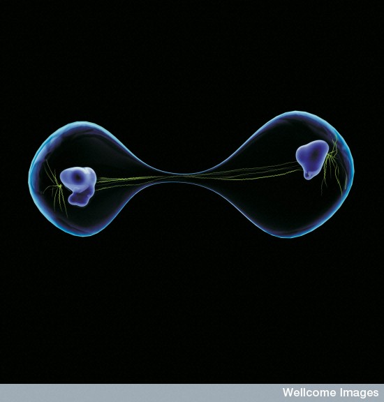
The study shows RhoA is necessary for proper regulation of a cellular process called cytokinesis during the final stage of cell division in hematopoietic progenitor cells, which produce specific types of blood cells.
Cytokinesis helps control the separation and grouping of genetic material as cells divide to decide their eventual fate.
Although the research was conducted in mouse models, the investigators feel their findings will be important to the future study of various blood diseases, immune disorders and cancers. The data could also be useful for research into prospective strategies for regenerative medicine, in which pluripotent stem cells could be used to attempt the repair or regrowth of damaged tissues.
One example is human combined immunodeficiency, which has been linked to mutations in the RhoA pathway. The immune disorder makes people highly susceptible to infections their body cannot fight and its underlying cause remains unclear.
"We show that RhoA deficiency causes hematopoietic failure in all lines of blood cells and results in defective hematopoietic progenitor cells," said Yi Zheng, PhD, lead investigator and director of Experimental Hematology and Cancer Biology at Cincinnati Children's. "This is also important to understanding diseases like pancytopenia, in which people don't produce enough mature red and white blood cells and platelets. In regenerative medicine, it appears RhoA function would need to be artfully controlled to obtain functional blood cells."
Zheng and his colleagues conducted a series of experiments to confirm their data. One initial test involved subjecting hematopoietic stem cells in the mouse bone marrow to stressful conditions known to stimulate the production of blood cells. During this experiment, the researchers noted the involvement of active RhoA signaling during hematopoietic progenitor cell formation.
The researchers then tested the hematopoietic stem and progenitor cells of mice in which the RhoA gene was knocked out, causing depletion of RhoA protein in those cells. In one experiment, RhoA-deficient stem cells were transplanted into another group of mice to see how well they would function.
Following the transplant of RhoA-deficient stem cells, researchers were surprised to learn that these stem cells produced new cells, but were unable to produce more differentiated blood cells (multi-potent hematopoietic progenitor cells).
Instead, researchers watched as the accumulation of progenitor cells grew into cells with more than one nucleus (multi-nucleated cells) that then failed to complete division and underwent cell death (programmed necrosis).
In a final experiment, researchers were able to restore the normal function of hematopoietic stem and progenitor cells by reconstituting RhoA into the cells which resulted in the production of multi-lineage red and white blood cells.
Zheng and his colleagues have studied disruptions in the RhoA GTPase pathway for the role these disruptions play in cancer formation, in which RhoA activity is often elevated.
The team has developed prospective small molecular inhibitors to block abnormal pathway functions as possible new targeted treatments for various cancers.
Zheng said researchers are using data from their previous work, along with the current study, to look for new strategies to fight various blood or immune system disorders.
Abstract
Hematopoietic progenitor cells (HPCs) are central to hematopoiesis as they provide large numbers of lineage-defined blood cells necessary to sustain blood homeostasis. They are one of the most actively cycling somatic cells, and their precise control is critical for hematopoietic homeostasis. The small GTPase RhoA is an intracellular molecular switch that integrates cytokine, chemokine, and adhesion signals to coordinate multiple context-dependent cellular processes. By using a RhoA conditional knockout mouse model, we show that RhoA deficiency causes a multilineage hematopoietic failure that is associated with defective multipotent HPCs. Interestingly, RhoA−/− hematopoietic stem cells retained long-term engraftment potential but failed to produce multipotent HPCs and lineage-defined blood cells. This multilineage hematopoietic failure was rescued by reconstituting wild-type RhoA into the RhoA−/− Lin−Sca-1+c-Kit+ compartment. Mechanistically, RhoA regulates actomyosin signaling, cytokinesis, and programmed necrosis of the HPCs, and loss of RhoA results in a cytokinesis failure of HPCs manifested by an accumulation of multinucleated cells caused by failed abscission of the cleavage furrow after telophase. Concomitantly, the HPCs show a drastically increased death associated with increased TNF–RIP-mediated necrosis. These results show that RhoA is a critical and specific regulator of multipotent HPCs during cytokinesis and thus essential for multilineage hematopoiesis.
Funding support for the study came in part from the National Institutes of Health (R01CA150547, P30DK090971, R01AG040118), the Deutsche Forschungsgemeinschaft (KFO142, GE2063/1), the Forschungsprogramm "Internationale Spitzenforschung II/3" der Baden-Württemberg Stiftung (P-BWS-SPII/3-06).
About Cincinnati Children's:
Cincinnati Children's Hospital Medical Center ranks third in the nation among all Honor Roll hospitals in U.S. News and World Report's 2013 Best Children's Hospitals ranking. It is ranked #1 for cancer and in the top 10 for nine of 10 pediatric specialties. Cincinnati Children's, a non-profit organization, is one of the top three recipients of pediatric research grants from the National Institutes of Health, and a research and teaching affiliate of the University of Cincinnati College of Medicine. The medical center is internationally recognized for improving child health and transforming delivery of care through fully integrated, globally recognized research, education and innovation. Additional information can be found at http://www.cincinnatichildrens.org. Connect on the Cincinnati Children's blog, via Facebook and on Twitter.
Original press releas: http://www.eurekalert.org/pub_releases/2013-10/cchm-sie100713.php
|