Healing powers
How do cells know to spread to cover and close a wound — or build an embryo? A team of researchers publishes new insights into how epithelial cells manage to spread out and in the case of a wound, cover an injury.
Spreading of the epithelial cell layer is fundamental for closure of wounds, as well as for embryonic development. The challenge is that the epithelial layer needs to increase in surface area, while maintaining its integrity.
Writing in the current online edition of Nature Cell Biology, The Institute of Science and Technology Austria (IST Austria) Professor Carl-Philipp Heisenberg, led a research team including first author Pedro Campinho, PhD student, in the exploration of epiboly in zebrafish development.
Epiboly is a step in the embryonic development of zebrafish when a thin epithelial cell layer spreads over the entire cell sphere within only 6 hours. This fast spreading comes along with a rapid increase in tension in the epithelium’s surface area. Researchers found they could orient the direction in which cells divide by controlling this tension.
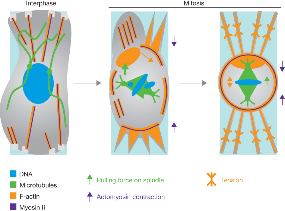
Controlling surface tension requires controlling two processes: (1) cell elongation and (2) the correct alignment of the mitotic spindle — the cell’s control center for division.
Tension in the epithelium elongates a cell, myosin II activity aligns the mitotic spindle with the axis of tension. Cells then divide perpendicular to the long axis.
In the absence of cell division, tissue tension increases. Researchers observed an ectopic fusion of cells as an alternative for tension release. They conclude that cell-division oriented by tension is key to ensuring tissue integrity and tissue spreading during epiboly.
Abstract
Epithelial spreading is a common and fundamental aspect of various developmental and disease-related processes such as epithelial closure and wound healing. A key challenge for epithelial tissues undergoing spreading is to increase their surface area without disrupting epithelial integrity. Here we show that orienting cell divisions by tension constitutes an efficient mechanism by which the enveloping cell layer (EVL) releases anisotropic tension while undergoing spreading during zebrafish epiboly. The control of EVL cell-division orientation by tension involves cell elongation and requires myosin II activity to align the mitotic spindle with the main tension axis. We also found that in the absence of tension-oriented cell divisions and in the presence of increased tissue tension, EVL cells undergo ectopic fusions, suggesting that the reduction of tension anisotropy by oriented cell divisions is required to prevent EVL cells from fusing. We conclude that cell-division orientation by tension constitutes a key mechanism for limiting tension anisotropy and thus promoting tissue spreading during EVL epiboly.
About IST Austria
The Institute of Science and Technology Austria (IST Austria) is a PhD granting institution located in the city of Klosterneuburg, 18 km from the center of Vienna. The Institute is dedicated to cutting-edge research in the natural, mathematical and computer sciences. Established jointly by the federal government of Austria and the provincial government of Lower Austria, the Institute was inaugurated in 2009 and will grow to about 100 research groups with 1000 scientists by 2026.
The governance and management structures of IST Austria guarantee its independence and freedom from political and commercial influences. The Institute is headed by the President, who is appointed by the Board of Trustees and advised by the Scientific Board. The first President of IST Austria is Thomas A. Henzinger, a leading computer scientist and former professor of the University of California at Berkeley and the EPFL Lausanne in Switzerland.
Original press release: http://ist.ac.at/en/news-media/news/news-detail/article/healing-powers/6/
|


 WOUND healing_CellBio.jpg)
 Related Article: