Welcome to The Visible Embryo


Home-- -History-- -Bibliography- -Pregnancy Timeline- --Prescription Drugs in Pregnancy- -- Pregnancy Calculator- --Female Reproductive System- -Contact
 

Welcome to The Visible Embryo, a comprehensive educational resource on human development from conception to birth.

The Visible Embryo provides visual references for changes in fetal development throughout pregnancy and can be navigated via fetal development or maternal changes.

The National Institutes of Child Health and Human Development awarded Phase I and Phase II Small Business Innovative Research Grants to develop The Visible Embryo. Initally designed to evaluate the internet as a teaching tool for first year medical students, The Visible Embryo is linked to over 600 educational institutions and is viewed by more than one million visitors each month.

Today, The Visible Embryo is linked to over 600 educational institutions and is viewed by more than 1 million visitors each month. The field of early embryology has grown to include the identification of the stem cell as not only critical to organogenesis in the embryo, but equally critical to organ function and repair in the adult human. The identification and understanding of genetic malfunction, inflammatory responses, and the progression in chronic disease, begins with a grounding in primary cellular and systemic functions manifested in the study of the early embryo.

WHO International Clinical Trials Registry Platform


The World Health Organization (WHO) has created a new Web site to help researchers, doctors and
patients obtain reliable information on high-quality clinical trials. Now you can go to one website and search all registers to identify clinical trial research underway around the world!


Home

History

Bibliography

Pregnancy Timeline

Prescription Drug Effects on Pregnancy

Pregnancy Calculator

Female Reproductive System

Contact The Visible Embryo

News Alerts Archive

Disclaimer: The Visible Embryo web site is provided for your general information only. The information contained on this site should not be treated as a substitute for medical, legal or other professional advice. Neither is The Visible Embryo responsible or liable for the contents of any websites of third parties which are listed on this site.
Content protected under a Creative Commons License.

No dirivative works may be made or used for commercial purposes.


Pregnancy Timeline by SemestersFetal liver is producing blood cellsHead may position into pelvisBrain convolutions beginFull TermWhite fat begins to be madeWhite fat begins to be madeHead may position into pelvisImmune system beginningImmune system beginningPeriod of rapid brain growthBrain convolutions beginLungs begin to produce surfactantSensory brain waves begin to activateSensory brain waves begin to activateInner Ear Bones HardenBone marrow starts making blood cellsBone marrow starts making blood cellsBrown fat surrounds lymphatic systemFetal sexual organs visibleFinger and toe prints appearFinger and toe prints appearHeartbeat can be detectedHeartbeat can be detectedBasic Brain Structure in PlaceThe Appearance of SomitesFirst Detectable Brain WavesA Four Chambered HeartBeginning Cerebral HemispheresFemale Reproductive SystemEnd of Embryonic PeriodEnd of Embryonic PeriodFirst Thin Layer of Skin AppearsThird TrimesterSecond TrimesterFirst TrimesterFertilizationDevelopmental Timeline
CLICK ON weeks 0 - 40 and follow along every 2 weeks of fetal development
Google Search artcles published since 2007


Home | Pregnancy Timeline | News Alerts |News Archive April 8, 2014


Components of a typical mitochondrion

1 Outer membrane: 1.1 Porin
2 Intermembrane space: 2.1 Intracristal space - 2.2 Peripheral space
3 Lamella: 3.1 Inner membrane - 3.11 Inner boundary membrane - 3.12 Cristal membrane - 3.2 Matrix - 3.3 Cristæ
4 Mitochondrial DNA
5 Matrix granule
6 Ribosome
7 ATP synthase

Image credit: www.Wikipedia.org


WHO Child Growth Charts

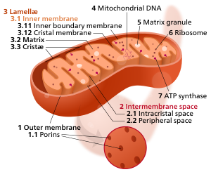
Potential treatment for mitochondrial disorders

Despite a fairly strong understanding of the pathology of some genetic mitochondrial disorders, efforts to treat them have been mostly ineffective.

Mitochondria — long known as "cellular power plants" because they generate the key energy source adenosine triphosphate (ATP) — are essential to proper cell function. Mitochondrial defects are often observed in diseases including cancer, Alzheimer's, and Parkinson's.

But now, graduate student Walter Chen and postdoctoral researcher Kivanc Birsoy, both in David Sabatini's lab at the Whitehead Institute, have unraveled how to rescue cells suffering from mitochondrial dysfunction. Chen and Birsoy suppressed mitochondrial function using the drug antimycin, in a test developed by Thijn Brummelkamp, former Whitehead Fellow. Through the test, Chen and Birsoy found that the gene ATPIF1 was protected against loss of mitochondrial function.

ATPIF1 is part of a backup system to save cells under conditions of starvation. When cells are deprived of oxygen and sugars, a mitochondrial complex called ATP synthase, switches from making to consuming ATP — harmful to an already starving cell. ATPIF1 interacts with ATP synthase, shutting it down to prevent it from consuming the mitochondria's dwindling ATP supply. But, the process worsens the mitochondrion's membrane potential


"In these diseases of mitochondrial dysfunction, in a sense, it's a false starvation situation for the cell—there are plenty of nutrients, but because there's a block in the mitochondria's normal function, the mitochondria behave as if there's not enough oxygen.

"So in these situations, activation of ATPIF1 is not good, because there are still many nutrients around to provide ATP. Instead, blocking ATPIF1 is therapeutic because it allows for maintenance of the membrane potential."

Walter Chen, postdoctoral researcher, the Whitehead Institute


Liver cells are frequently affected in patients with severe mitochondrial disease, so Chen and Birsoy tested the effects of mitochondrial dysfunction in the liver cells of control mice and mice with ATPIF1 genetically knocked out. Again, the liver cells with suppressed ATPIF1 function dealt better with mitochondrial dysfunction than liver cells with normal ATPIF1 activity.

"It's very simple—if you get rid of ATPIF1, you survive in the presence of mitochondrial dysfunction," says Birsoy. "From what we see so far, there are no major side effects from blocking ATPIF1 in mice."

For Chen and Birsoy, the next step in this line of research is to test the effects of ATPIF1 suppression in mouse models of mitochondrial dysfunction. Then they will try to identify therapeutics that effectively block ATPIF1 function.

The results are published in Cell Reports, April 10, 2014 as "Inhibition of ATPIF1 Ameliorates Severe Mitochondrial Respiratory Chain Dysfunction in Mammalian Cells."

Summary
Mitochondrial respiratory chain disorders are characterized by loss of electron transport chain (ETC) activity. Although the causes of many such diseases are known, there is a lack of effective therapies. To identify genes that confer resistance to severe ETC dysfunction when inactivated, we performed a genome-wide genetic screen in haploid human cells with the mitochondrial complex III inhibitor antimycin. This screen revealed that loss of ATPIF1 strongly protects against antimycin-induced ETC dysfunction and cell death by allowing for the maintenance of mitochondrial membrane potential. ATPIF1 loss protects against other forms of ETC dysfunction and is even essential for the viability of human ρ° cells lacking mitochondrial DNA, a system commonly used for studying ETC dysfunction. Importantly, inhibition of ATPIF1 ameliorates complex III blockade in primary hepatocytes, a cell type afflicted in severe mitochondrial disease. Altogether, these results suggest that inhibition of ATPIF1 can ameliorate severe ETC dysfunction in mitochondrial pathology.

This is an open access article under the CC BY license (http://creativecommons.org/licenses/by/3.0/).

This work is supported by National Institutes of Health (CA103866, CA129105, and AI07389), David H. Koch Institute for Integrative Cancer Research, Alexander and Margaret Stewart Trust Fund, National Institute of Aging, Jane Coffin Childs Memorial Fund, Leukemia and Lymphoma Society, and Damon Runyon Cancer Research Foundation.

David Sabatini's primary affiliation is with Whitehead Institute for Biomedical Research, where his laboratory is located and all his research is conducted. He is also a Howard Hughes Medical Institute investigator and a professor of biology at Massachusetts Institute of Technology.

Return to top of page