|
|
Welcome to The Visible Embryo, a comprehensive educational resource on human development from conception to birth.
The Visible Embryo provides visual references for changes in fetal development throughout pregnancy and can be navigated via fetal development or maternal changes.
The National Institutes of Child Health and Human Development awarded Phase I and Phase II Small Business Innovative Research Grants to develop The Visible Embryo. Initally designed to evaluate the internet as a teaching tool for first year medical students, The Visible Embryo is linked to over 600 educational institutions and is viewed by more than one million visitors each month.
Today, The Visible Embryo is linked to over 600 educational institutions and is viewed by more than 1 million visitors each month. The field of early embryology has grown to include the identification of the stem cell as not only critical to organogenesis in the embryo, but equally critical to organ function and repair in the adult human. The identification and understanding of genetic malfunction, inflammatory responses, and the progression in chronic disease, begins with a grounding in primary cellular and systemic functions manifested in the study of the early embryo.

The World Health Organization (WHO) has created a new Web site to help researchers, doctors and
patients obtain reliable information on high-quality clinical trials. Now you can go to one website and search all registers to identify clinical trial research underway around the world!
|
|
| Disclaimer: The Visible Embryo web site is provided for your general information only. The information contained on this site should not be treated as a substitute for medical, legal or other professional advice. Neither is The Visible Embryo responsible or liable for the contents of any websites of third parties which are listed on this site. |
|
|
 |
|
Content protected under a Creative Commons License. Commons License.
No dirivative works may be made or used for commercial purposes. |
|
|
| |
|
|

CLICK ON weeks 0 - 40 and follow along every 2 weeks of fetal development
|
|
|
|
|
Home | Pregnancy Timeline | News Alerts |News Archive May 2, 2014

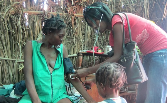
Haitian mother receiving midwife checkup.
Malnutrition in pregnancy can predispose a mother's newborn, her grand children,
great grandchildren and onwards, to metabolic disorders and disease.
|
|
 |
|
|
|
Pregnancy malnutrition effects last for generations
New research reveals environmental factors can predispose a mother's newborn, her grand children, great grandchildren and onwards, to metabolic disorders and disease.
In the journal Cell Metabolism researchers report that pregnant mice who are malnourished — having a 50% caloric restriction during the last week of pregnancy — bear pups who are low in birth weight, but then go on to become obese and diabetic as they age. In a domino effect, pups of growth-restricted males also inherit a predisposition to metabolic abnormalities.
To find out how these effects arise, leading author Dr. Josep Jiménez-Chillarón from the Hospital Sant Joan de Déu in Spain, investigated the patterns of LXR gene expression in mice, finding that in utero malnutrition of males influenced that gene's expression.
The LXR gene contributes to regulation of fat and cholesterol in the liver. According to Jiménez-Chillarón: "This may contribute, in part, to the transmission of diabetes risk from parents to offspring."
If these findings hold true for humans, what a woman eats while pregnant may have effects on health and disease in her future grandchildren. This opens up the possibility that a predisposition to some complex diseases might be inherited independent of your genetic DNA. But, Jiménez-Chillarón feels it is important not to"blame" one's parents (or even grandparents) for disease.
"Current beliefs are that the majority of epigenetic modifications to the sperm and egg are erased to avoid transmission of environmentally caused change. But our data suggests that a few environmentally induced epigenetic changes may be passed on and maintained in the next generation and onward.
"Our view is that we inherit some predisposition, but it is our own lifestyle that will determine whether inherited risk will truly translate into disease. Hence, a healthy lifestyle is the best way to prevent any potentially inherited or newly acquired obesity or diabetes predisposition."
Josep Jiménez-Chillarón, PhD, MD, Hospital Sant Joan de Déu, Spain
Highlights
• In utero undernutrition in F1 males programs liver FFA metabolism in the offspring
• Altered FFA metabolism in the F2 is explained, partly, by altered Lxra expression
• Altered Lxra expression can be attributed to changes in DNA methylation
• This epigenetic mark is already present in sperm from the progenitors, F1 male mice
Summary
Obesity and type 2 diabetes have a heritable component that is not attributable to genetic factors. Instead, epigenetic mechanisms may play a role. We have developed a mouse model of intrauterine growth restriction (IUGR) by in utero malnutrition. IUGR mice developed obesity and glucose intolerance with aging. Strikingly, offspring of IUGR male mice also developed glucose intolerance. Here, we show that in utero malnutrition of F1 males influenced the expression of lipogenic genes in livers of F2 mice, partly due to altered expression of Lxra. In turn, Lxra expression is attributed to altered DNA methylation of its 5′ UTR region. We found the same epigenetic signature in the sperm of their progenitors, F1 males. Our data indicate that in utero malnutrition results in epigenetic modifications in germ cells (F1) that are subsequently transmitted and maintained in somatic cells of the F2, thereby influencing health and disease risk of the offspring.
Cell Metabolism, Martinez et al.: "In utero undernutrition in male mice programs liver lipid metabolism in the second-generation offspring involving altered Lxra DNA methylation."
Return to top of page |
