Welcome to The Visible Embryo
  o
Home-- -History-- -Bibliography- -Pregnancy Timeline- --Prescription Drugs in Pregnancy- -- Pregnancy Calculator- --Female Reproductive System- -Contact

Welcome to The Visible Embryo, a comprehensive educational resource on human development from conception to birth.

The Visible Embryo provides visual references for changes in fetal development throughout pregnancy and can be navigated via fetal development or maternal changes.

The National Institutes of Child Health and Human Development awarded Phase I and Phase II Small Business Innovative Research Grants to develop The Visible Embryo. Initally designed to evaluate the internet as a teaching tool for first year medical students, The Visible Embryo is linked to over 600 educational institutions and is viewed by more than one million visitors each month.

Today, The Visible Embryo is linked to over 600 educational institutions and is viewed by more than 1 million visitors each month. The field of early embryology has grown to include the identification of the stem cell as not only critical to organogenesis in the embryo, but equally critical to organ function and repair in the adult human. The identification and understanding of genetic malfunction, inflammatory responses, and the progression in chronic disease, begins with a grounding in primary cellular and systemic functions manifested in the study of the early embryo.

WHO International Clinical Trials Registry Platform


The World Health Organization (WHO) has created a new Web site to help researchers, doctors and patients obtain reliable information on high-quality clinical trials. Now you can go to one website and search all registers to identify clinical trial research underway around the world!


Home

History

Bibliography

Pregnancy Timeline

Prescription Drug Effects on Pregnancy

Pregnancy Calculator

Female Reproductive System

Contact The Visible Embryo

News Alerts Archive

Disclaimer: The Visible Embryo web site is provided for your general information only. The information contained on this site should not be treated as a substitute for medical, legal or other professional advice. Neither is The Visible Embryo responsible or liable for the contents of any websites of third parties which are listed on this site.
Content protected under a Creative Commons License.

No dirivative works may be made or used for commercial purposes.


Pregnancy Timeline by SemestersLungs begin to produce surfactantImmune system beginningHead may position into pelvisFull TermPeriod of rapid brain growthWhite fat begins to be madeHead may position into pelvisWhite fat begins to be madeImmune system beginningBrain convolutions beginBrain convolutions beginFetal liver is producing blood cellsSensory brain waves begin to activateSensory brain waves begin to activateInner Ear Bones HardenBone marrow starts making blood cellsBone marrow starts making blood cellsBrown fat surrounds lymphatic systemFetal sexual organs visibleFinger and toe prints appearFinger and toe prints appearHeartbeat can be detectedHeartbeat can be detectedBasic Brain Structure in PlaceThe Appearance of SomitesFirst Detectable Brain WavesA Four Chambered HeartBeginning Cerebral HemispheresFemale Reproductive SystemEnd of Embryonic PeriodEnd of Embryonic PeriodFirst Thin Layer of Skin AppearsThird TrimesterSecond TrimesterFirst TrimesterFertilizationDevelopmental Timeline
CLICK ON weeks 0 - 40 and follow along every 2 weeks of fetal development
Google Search artcles published since 2007


Home | Pregnancy Timeline | News Alerts |News Archive Apr 28, 2015

A mitochondrion
Image Credit:American Association for the Advancement of Science


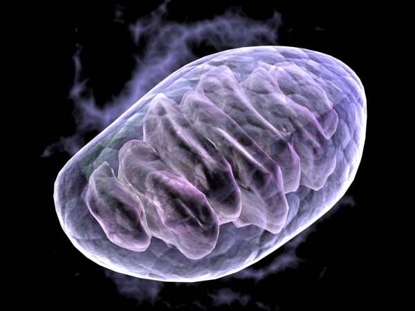
Mitochondrial genes and disease inheritance

Mitochondrial diseases are maternally inherited gene disorders that cause many debilitating conditions without any cures. Now, Salk Institute reports a successful gene-editing technology to prevent passing mutated mitochondrial DNA from mothers to offspring in mice.


"This technique is based on a single injection of mRNA into a mother's oocytes, or early embryos, and therefore could be easily implemented in IVF [in vitro fertilization] clinics throughout the world.

Since mutations in mitochondrial DNA have also been implicated in neurodegenerative disorders, cancer, and aging, our technology could potentially have broad clinical implications for preventing the transmission of disease-causing mutations to future generations."

Juan Carlos Izpisua Belmonte PhD, of the Salk Institute for Biological Studies and senior study author.


The study was published April 23 in the journal Cell.

Mitochondria are known as the powerhouses of the cell because they generate most of the cell's supply of ATP energy. ATP stands for an organic compound composed of adenosine (an adenine ring and a ribose sugar) and three phosphate groups. It releases energy when ATP is broken down (hydrolyzed) into ADP — Adenosine Diphosphate. The energy is used for many metabolic processes. So, ATP is considered the universal energy for metabolism. Each cell in the body contains anywhere from 1,000 to 100,000 copies of mitochondrial DNA, exclusively inherited from mom.

In most patients with mitochondrial disease, mutated and normal mitochondrial DNA molecules are mixed together. But, a high percentage of mutated mitochondrial DNA can lead to degeneration and catastrophic failure of various organs, resulting in serious health problems such as seizures, dementia, diabetes, heart failure, liver dysfunction, vision loss, and deafness.

Currently, therapies for preventing transmission of mitochondrial diseases from mother to child are limited. While genetic screening of embryos can partially reduce risk of transmitting mitochondrial diseases, another approach called "mitochondrial replacement therapy" actually transfers healthy mitochondria provided by a donor. This approach is being evaluated in the US, but it is soon to be allowed in the UK.


Mitochondrial replacement therapy has raised ethical, safety, and medical concerns because it involves combining genetic material from three different individuals: (1) the mother's egg, (2) the father's sperm, and (3) a donor's egg mitochondria.

Juan Carlos Izpisua Belmonte PhD


In the new study, Belmonte and his team demonstrated an alternative approach that allows for correction of the mutated DNA in mitochondria by using DNA-cutting enzymes called restriction endonucleases or TALENs. This gene-editing approach might be safer, simpler, and more ethical than mitochondrial replacement therapy because it does not require donor eggs. The enzymes are designed to target a specific mutated DNA sequence and introduce a precise cut that destroys the mutated mitochondrial DNA, while leaving normal mitochondrial DNA intact.

To test this approach, researchers created a mouse model carrying two specific types of mitochondrial DNA. TALENs were designed using restriction endonucleases to target and destroy only one of the DNA types. This approach decreased the levels of the targeted mitochondrial DNA, while sparing the untargeted mitochondrial DNA. The TALENs injected mouse embryos showed normal patterns of development, and were transferred to female mice. All pups born appeared healthy and with low levels of the targeted mitochondrial DNA in their various organs. They exhibited normal behavior, mitochondrial function, and genomic integrity. Even their offspring gave birth to pups that showed barely detectable levels of the targeted mitochondrial DNA. This first generation exercise demonstrates the effectiveness of this technique for preventing transgenerational transmission of mitochondrial diseases.

To confirm the clinical relevance of this strategy, researchers next screened and tested TALENs designed to target specific human mitochondrial DNA mutations. The specific disorders: (1) Leber's Hereditary Optic Neuropathy and Dystonia (LHOND) and (2) Neurogenic muscle weakness, Ataxia, and Retinitis Pigmentosa (NARP). This approach resulted in a significant reduction in mutated mitochondrial DNA in mouse eggs that contained genetic material from patient cells.


"We expect that this method will reduce the percentage of mutated mitochondrial DNA below the threshold for triggering mitochondrial diseases in humans."

Juan Carlos Izpisua Belmonte PhD


Before any clinical trials can begin, it will be necessary to evaluate the safety of the method on eggs from patients with mitochondrial diseases. Belmonte's team is collaborating with several IVF clinics to test the technology on surplus human eggs donated for research by patients with mitochondrial disease.

Abstract
Highlights
•Mitochondria-targeted nucleases selectively reduce mtDNA haplotypes in germline
•Germline heteroplasmy shift prevents transmission of mtDNA haplotypes to offspring
•Human mutated mtDNA can be reduced in oocytes by mitochondria-targeted nucleases

Summary
Mitochondrial diseases include a group of maternally inherited genetic disorders caused by mutations in mtDNA. In most of these patients, mutated mtDNA coexists with wild-type mtDNA, a situation known as mtDNA heteroplasmy. Here, we report on a strategy toward preventing germline transmission of mitochondrial diseases by inducing a mtDNA heteroplasmy shift through the selective elimination of mutated mtDNA. As a proof of concept, we took advantage of NZB/BALB heteroplasmic mice, which contain two mtDNA haplotypes, BALB and NZB, and selectively prevented their germline transmission using either mitochondria-targeted restriction endonucleases or TALENs. In addition, we successfully reduced human mutated mtDNA levels responsible for Leber’s hereditary optic neuropathy (LHOND), and neurogenic muscle weakness, ataxia, and retinitis pigmentosa (NARP), in mammalian oocytes using mitochondria-targeted TALEN (mito-TALENs). Our approaches represent a potential therapeutic avenue for preventing the transgenerational transmission of human mitochondrial diseases caused by mutations in mtDNA.

Cell, Reddy et al.: "Selective Elimination of Mitochondrial Mutations in the Germline by Genome Editing" http://dx.doi.org/10.1016/j.cell.2015.03.051

Cell, the flagship journal of Cell Press, is a bimonthly journal that publishes findings of unusual significance in any area of experimental biology, including but not limited to cell biology, molecular biology, neuroscience, immunology, virology and microbiology, cancer, human genetics, systems biology, signaling, and disease mechanisms and therapeutics. For more information, please visit http://www.cell.com/cell. To receive media alerts for Cell or other Cell Press journals, contact press@cell.com.

Return to top of page