|
|
Welcome to The Visible Embryo, a comprehensive educational resource on human development from conception to birth.
The Visible Embryo provides visual references for changes in fetal development throughout pregnancy and can be navigated via fetal development or maternal changes.
The National Institutes of Child Health and Human Development awarded Phase I and Phase II Small Business Innovative Research Grants to develop The Visible Embryo. Initally designed to evaluate the internet as a teaching tool for first year medical students, The Visible Embryo is linked to over 600 educational institutions and is viewed by more than one million visitors each month.
Today, The Visible Embryo is linked to over 600 educational institutions and is viewed by more than 1 million visitors each month. The field of early embryology has grown to include the identification of the stem cell as not only critical to organogenesis in the embryo, but equally critical to organ function and repair in the adult human. The identification and understanding of genetic malfunction, inflammatory responses, and the progression in chronic disease, begins with a grounding in primary cellular and systemic functions manifested in the study of the early embryo.

The World Health Organization (WHO) has created a new Web site to help researchers, doctors and patients obtain reliable information on high-quality clinical trials. Now you can go to one website and search all registers to identify clinical trial research underway around the world!

|
|
| Disclaimer: The Visible Embryo web site is provided for your general information only. The information contained on this site should not be treated as a substitute for medical, legal or other professional advice. Neither is The Visible Embryo responsible or liable for the contents of any websites of third parties which are listed on this site. |
|
|
 |
|
Content protected under a Creative Commons License. Commons License.
No dirivative works may be made or used for commercial purposes. |
|
|
| |
|
|

CLICK ON weeks 0 - 40 and follow along every 2 weeks of fetal development
|
|
|
|
|
Home | Pregnancy Timeline | News Alerts |News Archive May 13, 2015
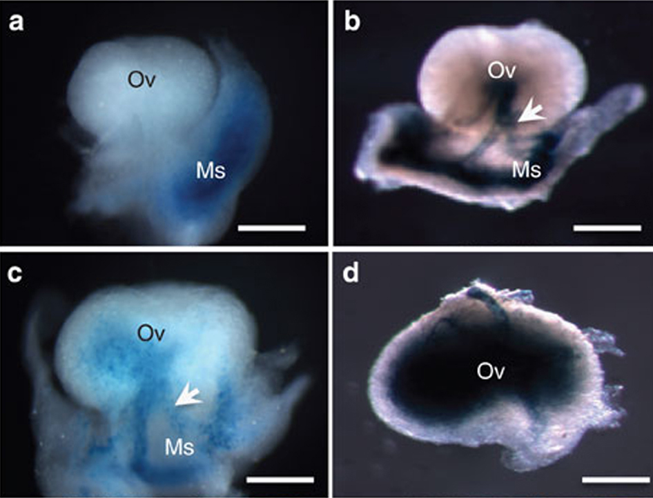
Gli1-LacZ in mouse ovaries is detected by staining for β-galactosidase (a–d)
Ov - Ovary; Ms - Mesenchyme. Arrow indicates mesonephric tubules.
Scale bar: a–d, 500 μm..
Image Credit: Nature Communications
|
|
|
|
|
|
Shedding new light on reproductive disorders
A National Institutes of Health study has solved an ovarian cell mystery. Scientists have figured out the origin of one of the cell types that make up the ovary. They also discovered how ovarian cells share data when a developing follicle holds the next maturing egg.
Researchers believe this new information will help them understand the cause of ovarian disorders such as premature ovary failure and polycystic ovarian syndrome, both conditions result in hormone imbalances and impact fertility in women.
Researchers at the National Institute of Environmental Health Sciences (NIEHS) published the findings online April 28 in the journal Nature Communications. According to NIEHS researcher and corresponding author Humphrey Yao PhD, the ovarian follicle is the basic functional unit of the ovary. It contains the egg surrounded by two distinct cell types, known as granulosa cells and theca cells. Scientists have known the cellular origins of the egg and granulosa cells, but not where theca cells came from or what directs their development.
Yao: "This question remained unanswered for decades, but using a technique called lineage tracing, we determined that theca cells in mice come from both inside and outside the ovary, from embryonic tissue called mesenchyme."
Without theca cells, women cannot produce the hormones that sustain follicle growth. However, one of the major hormones theca cells produce is androgen, widely thought of as a male hormone. But in a superb example of teamwork, granulosa cells convert androgen to estrogen.
As a result of their investigation, Yao and his colleagues were able to trace molecular signals from theca cells all the way to androgen production. The pathway is triggered when theca cells signal granulosa cells to stimulate the oocyte or immature egg to mature. Being able to trace this pathway gives scientists insight to when and how particular ovarian disorders might arise.
According to Chang Liu PhD, a visiting fellow in Yao's group and first author on the paper: "The problem starts within the theca cell compartment... [knowing] what makes these cells grow, we can search for possible genetic mutations or environmental factors that affect the process leading to ovarian cell disorders."
Yao wants to continue exploring the two cell types sourcing theca cells. And as his research has been carried out in mice, he must verify how the communication pathway is or is not the same in humans. Imbalances or malfunctions along the pathway have the potential to reveal reasons behind some failures in female fertility.
Abstract
Organogenesis of the ovary is a highly orchestrated process involving multiple lineage determination of ovarian surface epithelium, granulosa cells and theca cells. Although the sources of ovarian surface epithelium and granulosa cells are known, the origin(s) of theca progenitor cells have not been definitively identified. Here we show that theca cells derive from two sources: Wt1+ cells indigenous to the ovary and Gli1+ mesenchymal cells that migrate from the mesonephros. These progenitors acquire theca lineage marker Gli1 in response to paracrine signals Desert hedgehog (Dhh) and Indian hedgehog (Ihh) from granulosa cells. Ovaries lacking Dhh/Ihh exhibit theca layer loss, blunted steroid production, arrested folliculogenesis and failure to form corpora lutea. Production of Dhh/Ihh in granulosa cells requires growth differentiation factor 9 (GDF9) from the oocyte. Our studies provide the first genetic evidence for the origins of theca cells and reveal a multicellular interaction critical for the formation of a functional theca.
NIEHS supports research to understand the effects of the environment on human health and is part of NIH. For more information on environmental health topics, visit http://www.niehs.nih.gov. Subscribe to one or more of the NIEHS news lists to stay current on NIEHS news, press releases, grant opportunities, training, events, and publications.
About the National Institutes of Health (NIH): NIH, the nation's medical research agency, includes 27 Institutes and Centers and is a component of the U.S. Department of Health and Human Services. NIH is the primary federal agency conducting and supporting basic, clinical, and translational medical research, and is investigating the causes, treatments, and cures for both common and rare diseases. For more information about NIH and its programs, visit http://www.nih.gov.
Return to top of page
|